Maariyah Ahmed, Myra Garzanich, Luigi E Melaragno, Sarah Nyirjesy, Natalia Von Windheim, Matthew Marquardt, Michael Luttrull, Nathan Quails, Kyle K VanKoevering
{"title":"探索 CT 像素和体素大小对下颌骨重建中解剖建模的影响。","authors":"Maariyah Ahmed, Myra Garzanich, Luigi E Melaragno, Sarah Nyirjesy, Natalia Von Windheim, Matthew Marquardt, Michael Luttrull, Nathan Quails, Kyle K VanKoevering","doi":"10.1186/s41205-024-00223-0","DOIUrl":null,"url":null,"abstract":"<p><strong>Background: </strong>Computer-aided modeling and design (CAM/CAD) of patient anatomy from computed tomography (CT) imaging and 3D printing technology enable the creation of tangible, patient-specific anatomic models that can be used for surgical guidance. These models have been associated with better patient outcomes; however, a lack of CT imaging guidelines risks the capture of unsuitable imaging for patient-specific modeling. This study aims to investigate how CT image pixel size (X-Y) and slice thickness (Z) impact the accuracy of mandibular models.</p><p><strong>Methods: </strong>Six cadaver heads were CT scanned at varying slice thicknesses and pixel sizes and turned into CAD models of the mandible for each scan. The cadaveric mandibles were then dissected and surface scanned, producing a CAD model of the true anatomy to be used as the gold standard for digital comparison. The root mean square (RMS) value of these comparisons, and the percentage of points that deviated from the true cadaveric anatomy by over 2.00 mm were used to evaluate accuracy. Two-way ANOVA and Tukey-Kramer post-hoc tests were used to determine significant differences in accuracy.</p><p><strong>Results: </strong>Two-way ANOVA demonstrated significant difference in RMS for slice thickness but not pixel size while post-hoc testing showed a significant difference in pixel size only between pixels of 0.32 mm and 1.32 mm. For slice thickness, post-hoc testing revealed significantly smaller RMS values for scans with slice thicknesses of 0.67 mm, 1.25 mm, and 3.00 mm compared to those with a slice thickness of 5.00 mm. No significant differences were found between 0.67 mm, 1.25 mm, and 3.00 mm slice thicknesses. Results for the percentage of points deviating from cadaveric anatomy greater than 2.00 mm agreed with those for RMS except when comparing pixel sizes of 0.75 mm and 0.818 mm against 1.32 mm in post-hoc testing, which showed a significant difference as well.</p><p><strong>Conclusion: </strong>This study suggests that slice thickness has a more significant impact on 3D model accuracy than pixel size, providing objective validation for guidelines favoring rigorous standards for slice thickness while recommending isotropic voxels. Additionally, our results indicate that CT scans up to 3.00 mm in slice thickness may provide an adequate 3D model for facial bony anatomy, such as the mandible, depending on the clinical indication.</p>","PeriodicalId":72036,"journal":{"name":"3D printing in medicine","volume":"10 1","pages":"21"},"PeriodicalIF":3.1000,"publicationDate":"2024-06-26","publicationTypes":"Journal Article","fieldsOfStudy":null,"isOpenAccess":false,"openAccessPdf":"https://www.ncbi.nlm.nih.gov/pmc/articles/PMC11202317/pdf/","citationCount":"0","resultStr":"{\"title\":\"Exploring CT pixel and voxel size effect on anatomic modeling in mandibular reconstruction.\",\"authors\":\"Maariyah Ahmed, Myra Garzanich, Luigi E Melaragno, Sarah Nyirjesy, Natalia Von Windheim, Matthew Marquardt, Michael Luttrull, Nathan Quails, Kyle K VanKoevering\",\"doi\":\"10.1186/s41205-024-00223-0\",\"DOIUrl\":null,\"url\":null,\"abstract\":\"<p><strong>Background: </strong>Computer-aided modeling and design (CAM/CAD) of patient anatomy from computed tomography (CT) imaging and 3D printing technology enable the creation of tangible, patient-specific anatomic models that can be used for surgical guidance. These models have been associated with better patient outcomes; however, a lack of CT imaging guidelines risks the capture of unsuitable imaging for patient-specific modeling. This study aims to investigate how CT image pixel size (X-Y) and slice thickness (Z) impact the accuracy of mandibular models.</p><p><strong>Methods: </strong>Six cadaver heads were CT scanned at varying slice thicknesses and pixel sizes and turned into CAD models of the mandible for each scan. The cadaveric mandibles were then dissected and surface scanned, producing a CAD model of the true anatomy to be used as the gold standard for digital comparison. The root mean square (RMS) value of these comparisons, and the percentage of points that deviated from the true cadaveric anatomy by over 2.00 mm were used to evaluate accuracy. Two-way ANOVA and Tukey-Kramer post-hoc tests were used to determine significant differences in accuracy.</p><p><strong>Results: </strong>Two-way ANOVA demonstrated significant difference in RMS for slice thickness but not pixel size while post-hoc testing showed a significant difference in pixel size only between pixels of 0.32 mm and 1.32 mm. For slice thickness, post-hoc testing revealed significantly smaller RMS values for scans with slice thicknesses of 0.67 mm, 1.25 mm, and 3.00 mm compared to those with a slice thickness of 5.00 mm. No significant differences were found between 0.67 mm, 1.25 mm, and 3.00 mm slice thicknesses. Results for the percentage of points deviating from cadaveric anatomy greater than 2.00 mm agreed with those for RMS except when comparing pixel sizes of 0.75 mm and 0.818 mm against 1.32 mm in post-hoc testing, which showed a significant difference as well.</p><p><strong>Conclusion: </strong>This study suggests that slice thickness has a more significant impact on 3D model accuracy than pixel size, providing objective validation for guidelines favoring rigorous standards for slice thickness while recommending isotropic voxels. Additionally, our results indicate that CT scans up to 3.00 mm in slice thickness may provide an adequate 3D model for facial bony anatomy, such as the mandible, depending on the clinical indication.</p>\",\"PeriodicalId\":72036,\"journal\":{\"name\":\"3D printing in medicine\",\"volume\":\"10 1\",\"pages\":\"21\"},\"PeriodicalIF\":3.1000,\"publicationDate\":\"2024-06-26\",\"publicationTypes\":\"Journal Article\",\"fieldsOfStudy\":null,\"isOpenAccess\":false,\"openAccessPdf\":\"https://www.ncbi.nlm.nih.gov/pmc/articles/PMC11202317/pdf/\",\"citationCount\":\"0\",\"resultStr\":null,\"platform\":\"Semanticscholar\",\"paperid\":null,\"PeriodicalName\":\"3D printing in medicine\",\"FirstCategoryId\":\"1085\",\"ListUrlMain\":\"https://doi.org/10.1186/s41205-024-00223-0\",\"RegionNum\":0,\"RegionCategory\":null,\"ArticlePicture\":[],\"TitleCN\":null,\"AbstractTextCN\":null,\"PMCID\":null,\"EPubDate\":\"\",\"PubModel\":\"\",\"JCR\":\"Q1\",\"JCRName\":\"RADIOLOGY, NUCLEAR MEDICINE & MEDICAL IMAGING\",\"Score\":null,\"Total\":0}","platform":"Semanticscholar","paperid":null,"PeriodicalName":"3D printing in medicine","FirstCategoryId":"1085","ListUrlMain":"https://doi.org/10.1186/s41205-024-00223-0","RegionNum":0,"RegionCategory":null,"ArticlePicture":[],"TitleCN":null,"AbstractTextCN":null,"PMCID":null,"EPubDate":"","PubModel":"","JCR":"Q1","JCRName":"RADIOLOGY, NUCLEAR MEDICINE & MEDICAL IMAGING","Score":null,"Total":0}
Exploring CT pixel and voxel size effect on anatomic modeling in mandibular reconstruction.
Background: Computer-aided modeling and design (CAM/CAD) of patient anatomy from computed tomography (CT) imaging and 3D printing technology enable the creation of tangible, patient-specific anatomic models that can be used for surgical guidance. These models have been associated with better patient outcomes; however, a lack of CT imaging guidelines risks the capture of unsuitable imaging for patient-specific modeling. This study aims to investigate how CT image pixel size (X-Y) and slice thickness (Z) impact the accuracy of mandibular models.
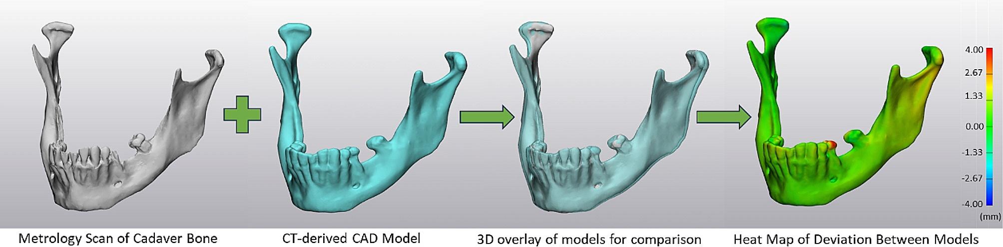
Methods: Six cadaver heads were CT scanned at varying slice thicknesses and pixel sizes and turned into CAD models of the mandible for each scan. The cadaveric mandibles were then dissected and surface scanned, producing a CAD model of the true anatomy to be used as the gold standard for digital comparison. The root mean square (RMS) value of these comparisons, and the percentage of points that deviated from the true cadaveric anatomy by over 2.00 mm were used to evaluate accuracy. Two-way ANOVA and Tukey-Kramer post-hoc tests were used to determine significant differences in accuracy.
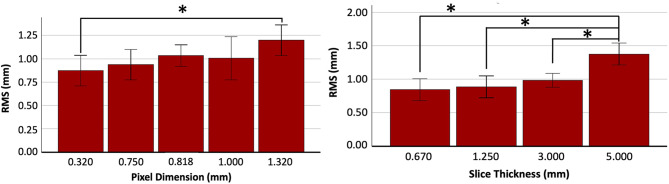
Results: Two-way ANOVA demonstrated significant difference in RMS for slice thickness but not pixel size while post-hoc testing showed a significant difference in pixel size only between pixels of 0.32 mm and 1.32 mm. For slice thickness, post-hoc testing revealed significantly smaller RMS values for scans with slice thicknesses of 0.67 mm, 1.25 mm, and 3.00 mm compared to those with a slice thickness of 5.00 mm. No significant differences were found between 0.67 mm, 1.25 mm, and 3.00 mm slice thicknesses. Results for the percentage of points deviating from cadaveric anatomy greater than 2.00 mm agreed with those for RMS except when comparing pixel sizes of 0.75 mm and 0.818 mm against 1.32 mm in post-hoc testing, which showed a significant difference as well.
Conclusion: This study suggests that slice thickness has a more significant impact on 3D model accuracy than pixel size, providing objective validation for guidelines favoring rigorous standards for slice thickness while recommending isotropic voxels. Additionally, our results indicate that CT scans up to 3.00 mm in slice thickness may provide an adequate 3D model for facial bony anatomy, such as the mandible, depending on the clinical indication.


 分享
分享

 求助内容:
求助内容: 应助结果提醒方式:
 扫码关注我们
