{"title":"ATAD3 是白色脂肪细胞样 3T3-L1 细胞线粒体生物生成和脂肪生成的限制因子。","authors":"Shuijie Li, Rui Xu, Yao Yao, Denis Rousseau","doi":"10.1002/cbin.12206","DOIUrl":null,"url":null,"abstract":"<p>ATAD3 is a vital ATPase of the inner mitochondrial membrane of pluri-cellular eukaryotes, with largely unknown functions but early required for organism development as necessary for mitochondrial biogenesis. ATAD3 knock-down in <i>C. elegans</i> inhibits at first the development of adipocyte-like intestinal tissue so we used mouse adipocyte model 3T3-L1 cells to analyze ATAD3 functions during adipogenesis and lipogenesis in a mammalian model. ATAD3 function was studied by stable and transient modulation of ATAD3 expression in adipogenesis- induced 3T3-L1 cells using Knock-Down and overexpression strategies, exploring different steps of adipocyte differentiation and lipogenesis. We show that (i) an increase in ATAD3 is preceding differentiation-induced mitochondrial biogenesis; (ii) downregulation of ATAD3 inhibits adipogenesis, lipogenesis, and impedes overexpression of many mitochondrial proteins; (iii) ATAD3 re-expression rescues the phenotype of ATAD3 KD, and (iv) differentiation and lipogenesis are accelerated by ATAD3 overexpression, but inhibited by expression of a dominant-negative mutant. We further show that the ATAD3 KD phenotype is not due to altered insulin signal but involves a limitation of mitochondrial biogenesis linked to Drp1. These results demonstrate that ATAD3 is limiting for in vitro mitochondrial biogenesis and adipogenesis/lipogenesis and therefore that ATAD3 mutation/over- or under-expression could be involved in adipogenic and lipogenic pathologies.</p>","PeriodicalId":9806,"journal":{"name":"Cell Biology International","volume":"48 10","pages":"1473-1489"},"PeriodicalIF":3.5000,"publicationDate":"2024-06-23","publicationTypes":"Journal Article","fieldsOfStudy":null,"isOpenAccess":false,"openAccessPdf":"https://onlinelibrary.wiley.com/doi/epdf/10.1002/cbin.12206","citationCount":"0","resultStr":"{\"title\":\"ATAD3 is a limiting factor in mitochondrial biogenesis and adipogenesis of white adipocyte-like 3T3-L1 cells\",\"authors\":\"Shuijie Li, Rui Xu, Yao Yao, Denis Rousseau\",\"doi\":\"10.1002/cbin.12206\",\"DOIUrl\":null,\"url\":null,\"abstract\":\"<p>ATAD3 is a vital ATPase of the inner mitochondrial membrane of pluri-cellular eukaryotes, with largely unknown functions but early required for organism development as necessary for mitochondrial biogenesis. ATAD3 knock-down in <i>C. elegans</i> inhibits at first the development of adipocyte-like intestinal tissue so we used mouse adipocyte model 3T3-L1 cells to analyze ATAD3 functions during adipogenesis and lipogenesis in a mammalian model. ATAD3 function was studied by stable and transient modulation of ATAD3 expression in adipogenesis- induced 3T3-L1 cells using Knock-Down and overexpression strategies, exploring different steps of adipocyte differentiation and lipogenesis. We show that (i) an increase in ATAD3 is preceding differentiation-induced mitochondrial biogenesis; (ii) downregulation of ATAD3 inhibits adipogenesis, lipogenesis, and impedes overexpression of many mitochondrial proteins; (iii) ATAD3 re-expression rescues the phenotype of ATAD3 KD, and (iv) differentiation and lipogenesis are accelerated by ATAD3 overexpression, but inhibited by expression of a dominant-negative mutant. We further show that the ATAD3 KD phenotype is not due to altered insulin signal but involves a limitation of mitochondrial biogenesis linked to Drp1. These results demonstrate that ATAD3 is limiting for in vitro mitochondrial biogenesis and adipogenesis/lipogenesis and therefore that ATAD3 mutation/over- or under-expression could be involved in adipogenic and lipogenic pathologies.</p>\",\"PeriodicalId\":9806,\"journal\":{\"name\":\"Cell Biology International\",\"volume\":\"48 10\",\"pages\":\"1473-1489\"},\"PeriodicalIF\":3.5000,\"publicationDate\":\"2024-06-23\",\"publicationTypes\":\"Journal Article\",\"fieldsOfStudy\":null,\"isOpenAccess\":false,\"openAccessPdf\":\"https://onlinelibrary.wiley.com/doi/epdf/10.1002/cbin.12206\",\"citationCount\":\"0\",\"resultStr\":null,\"platform\":\"Semanticscholar\",\"paperid\":null,\"PeriodicalName\":\"Cell Biology International\",\"FirstCategoryId\":\"99\",\"ListUrlMain\":\"https://onlinelibrary.wiley.com/doi/10.1002/cbin.12206\",\"RegionNum\":3,\"RegionCategory\":\"生物学\",\"ArticlePicture\":[],\"TitleCN\":null,\"AbstractTextCN\":null,\"PMCID\":null,\"EPubDate\":\"\",\"PubModel\":\"\",\"JCR\":\"Q3\",\"JCRName\":\"CELL BIOLOGY\",\"Score\":null,\"Total\":0}","platform":"Semanticscholar","paperid":null,"PeriodicalName":"Cell Biology International","FirstCategoryId":"99","ListUrlMain":"https://onlinelibrary.wiley.com/doi/10.1002/cbin.12206","RegionNum":3,"RegionCategory":"生物学","ArticlePicture":[],"TitleCN":null,"AbstractTextCN":null,"PMCID":null,"EPubDate":"","PubModel":"","JCR":"Q3","JCRName":"CELL BIOLOGY","Score":null,"Total":0}
ATAD3 is a limiting factor in mitochondrial biogenesis and adipogenesis of white adipocyte-like 3T3-L1 cells
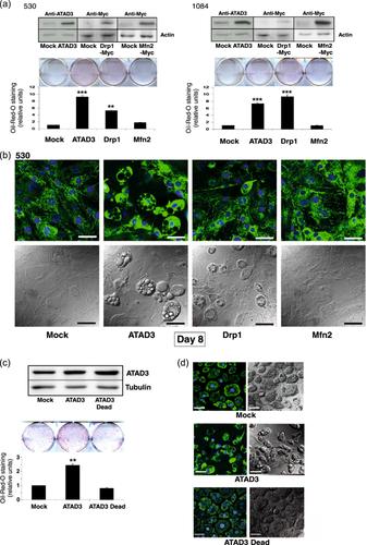
ATAD3 is a vital ATPase of the inner mitochondrial membrane of pluri-cellular eukaryotes, with largely unknown functions but early required for organism development as necessary for mitochondrial biogenesis. ATAD3 knock-down in C. elegans inhibits at first the development of adipocyte-like intestinal tissue so we used mouse adipocyte model 3T3-L1 cells to analyze ATAD3 functions during adipogenesis and lipogenesis in a mammalian model. ATAD3 function was studied by stable and transient modulation of ATAD3 expression in adipogenesis- induced 3T3-L1 cells using Knock-Down and overexpression strategies, exploring different steps of adipocyte differentiation and lipogenesis. We show that (i) an increase in ATAD3 is preceding differentiation-induced mitochondrial biogenesis; (ii) downregulation of ATAD3 inhibits adipogenesis, lipogenesis, and impedes overexpression of many mitochondrial proteins; (iii) ATAD3 re-expression rescues the phenotype of ATAD3 KD, and (iv) differentiation and lipogenesis are accelerated by ATAD3 overexpression, but inhibited by expression of a dominant-negative mutant. We further show that the ATAD3 KD phenotype is not due to altered insulin signal but involves a limitation of mitochondrial biogenesis linked to Drp1. These results demonstrate that ATAD3 is limiting for in vitro mitochondrial biogenesis and adipogenesis/lipogenesis and therefore that ATAD3 mutation/over- or under-expression could be involved in adipogenic and lipogenic pathologies.
期刊介绍:
Each month, the journal publishes easy-to-assimilate, up-to-the minute reports of experimental findings by researchers using a wide range of the latest techniques. Promoting the aims of cell biologists worldwide, papers reporting on structure and function - especially where they relate to the physiology of the whole cell - are strongly encouraged. Molecular biology is welcome, as long as articles report findings that are seen in the wider context of cell biology. In covering all areas of the cell, the journal is both appealing and accessible to a broad audience. Authors whose papers do not appeal to cell biologists in general because their topic is too specialized (e.g. infectious microbes, protozoology) are recommended to send them to more relevant journals. Papers reporting whole animal studies or work more suited to a medical journal, e.g. histopathological studies or clinical immunology, are unlikely to be accepted, unless they are fully focused on some important cellular aspect.
These last remarks extend particularly to papers on cancer. Unless firmly based on some deeper cellular or molecular biological principle, papers that are highly specialized in this field, with limited appeal to cell biologists at large, should be directed towards journals devoted to cancer, there being very many from which to choose.


 分享
分享
 求助内容:
求助内容: 应助结果提醒方式:
 扫码关注我们
