{"title":"NADPH 会改变 DUOX1 的钙响应性","authors":"Gregory E. Conner","doi":"10.1016/j.redox.2024.103251","DOIUrl":null,"url":null,"abstract":"<div><p>Hydrogen peroxide is a key element in redox signaling and in setting cellular redox tone. DUOX1 and DUOX2, that directly synthesize hydrogen peroxide, are the most abundant NADPH oxidase transcripts in most epithelia. DUOX1 and DUOX2 hydrogen peroxide synthesis is regulated by intracellular calcium transients and thus cells can respond to signals and initiate responses by increasing cellular hydrogen peroxide synthesis. Nevertheless, many details of their enzymatic regulation are still unexplored. DUOX1 and DUOXA1 were expressed in HEK293T cells and activity was studied in homogenates and membrane fractions. When DUOX1 homogenates or membranes were pre-incubated in NADPH and started with addition of Ca<sup>2+</sup>, to mimic intracellular activation, progress curves were distinctly different from those pre-incubated in Ca<sup>2+</sup> and started with NADPH. The Ca<sup>2+</sup> EC<sub>50</sub> for DUOX1's initial rate when pre-incubated in Ca<sup>2+</sup>, was three orders of magnitude lower (EC<sub>50</sub> ∼ 10<sup>−6</sup> M) than with preincubation in NADPH (EC<sub>50</sub> ∼ 10<sup>−3</sup> M). In addition, activity was several fold lower with Ca<sup>2+</sup> start. Identical results were obtained using homogenates and membrane fractions. The data suggested that DUOX1 Ca<sup>2+</sup> binding in expected physiological signaling conditions only slowly leads to maximal hydrogen peroxide synthesis and that full hydrogen peroxide synthesis activity <em>in vivo</em> only can occur when encountering extremely high concentration Ca<sup>2+</sup> signals. Thus, a complex interplay of intracellular NADPH and Ca<sup>2+</sup> concentrations regulate DUOX1 over a wide extent and may limit DUOX1 activity to a restricted range and spatial distribution.</p></div>","PeriodicalId":20998,"journal":{"name":"Redox Biology","volume":"75 ","pages":"Article 103251"},"PeriodicalIF":11.9000,"publicationDate":"2024-09-01","publicationTypes":"Journal Article","fieldsOfStudy":null,"isOpenAccess":false,"openAccessPdf":"https://www.sciencedirect.com/science/article/pii/S2213231724002295/pdfft?md5=5422e46303159707a7adb8e6d99dd1d8&pid=1-s2.0-S2213231724002295-main.pdf","citationCount":"0","resultStr":"{\"title\":\"NADPH Alters DUOX1 Calcium Responsiveness\",\"authors\":\"Gregory E. Conner\",\"doi\":\"10.1016/j.redox.2024.103251\",\"DOIUrl\":null,\"url\":null,\"abstract\":\"<div><p>Hydrogen peroxide is a key element in redox signaling and in setting cellular redox tone. DUOX1 and DUOX2, that directly synthesize hydrogen peroxide, are the most abundant NADPH oxidase transcripts in most epithelia. DUOX1 and DUOX2 hydrogen peroxide synthesis is regulated by intracellular calcium transients and thus cells can respond to signals and initiate responses by increasing cellular hydrogen peroxide synthesis. Nevertheless, many details of their enzymatic regulation are still unexplored. DUOX1 and DUOXA1 were expressed in HEK293T cells and activity was studied in homogenates and membrane fractions. When DUOX1 homogenates or membranes were pre-incubated in NADPH and started with addition of Ca<sup>2+</sup>, to mimic intracellular activation, progress curves were distinctly different from those pre-incubated in Ca<sup>2+</sup> and started with NADPH. The Ca<sup>2+</sup> EC<sub>50</sub> for DUOX1's initial rate when pre-incubated in Ca<sup>2+</sup>, was three orders of magnitude lower (EC<sub>50</sub> ∼ 10<sup>−6</sup> M) than with preincubation in NADPH (EC<sub>50</sub> ∼ 10<sup>−3</sup> M). In addition, activity was several fold lower with Ca<sup>2+</sup> start. Identical results were obtained using homogenates and membrane fractions. The data suggested that DUOX1 Ca<sup>2+</sup> binding in expected physiological signaling conditions only slowly leads to maximal hydrogen peroxide synthesis and that full hydrogen peroxide synthesis activity <em>in vivo</em> only can occur when encountering extremely high concentration Ca<sup>2+</sup> signals. Thus, a complex interplay of intracellular NADPH and Ca<sup>2+</sup> concentrations regulate DUOX1 over a wide extent and may limit DUOX1 activity to a restricted range and spatial distribution.</p></div>\",\"PeriodicalId\":20998,\"journal\":{\"name\":\"Redox Biology\",\"volume\":\"75 \",\"pages\":\"Article 103251\"},\"PeriodicalIF\":11.9000,\"publicationDate\":\"2024-09-01\",\"publicationTypes\":\"Journal Article\",\"fieldsOfStudy\":null,\"isOpenAccess\":false,\"openAccessPdf\":\"https://www.sciencedirect.com/science/article/pii/S2213231724002295/pdfft?md5=5422e46303159707a7adb8e6d99dd1d8&pid=1-s2.0-S2213231724002295-main.pdf\",\"citationCount\":\"0\",\"resultStr\":null,\"platform\":\"Semanticscholar\",\"paperid\":null,\"PeriodicalName\":\"Redox Biology\",\"FirstCategoryId\":\"99\",\"ListUrlMain\":\"https://www.sciencedirect.com/science/article/pii/S2213231724002295\",\"RegionNum\":1,\"RegionCategory\":\"生物学\",\"ArticlePicture\":[],\"TitleCN\":null,\"AbstractTextCN\":null,\"PMCID\":null,\"EPubDate\":\"2024/6/20 0:00:00\",\"PubModel\":\"Epub\",\"JCR\":\"Q1\",\"JCRName\":\"BIOCHEMISTRY & MOLECULAR BIOLOGY\",\"Score\":null,\"Total\":0}","platform":"Semanticscholar","paperid":null,"PeriodicalName":"Redox Biology","FirstCategoryId":"99","ListUrlMain":"https://www.sciencedirect.com/science/article/pii/S2213231724002295","RegionNum":1,"RegionCategory":"生物学","ArticlePicture":[],"TitleCN":null,"AbstractTextCN":null,"PMCID":null,"EPubDate":"2024/6/20 0:00:00","PubModel":"Epub","JCR":"Q1","JCRName":"BIOCHEMISTRY & MOLECULAR BIOLOGY","Score":null,"Total":0}
引用次数: 0
摘要
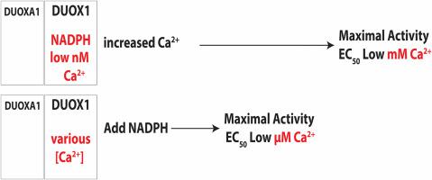
过氧化氢是氧化还原信号传递和确定细胞氧化还原基调的关键因素。直接合成过氧化氢的 DUOX1 和 DUOX2 是大多数上皮细胞中最丰富的 NADPH 氧化酶转录本。DUOX1 和 DUOX2 的过氧化氢合成受细胞内钙瞬态的调节,因此细胞可以通过增加细胞过氧化氢的合成来响应信号和启动反应。然而,它们的酶调控的许多细节仍有待探索。我们在 HEK293T 细胞中表达了 DUOX1 和 DUOXA1,并研究了它们在匀浆和膜分馏物中的活性。当 DUOX1 的匀浆或膜预先在 NADPH 中孵育并开始加入 Ca 以模拟细胞内活化时,其进展曲线与预先在 Ca 中孵育并开始加入 NADPH 的进展曲线明显不同。在 Ca 中预孵育时,DUOX1 初始速率的 Ca EC(EC ∼ 10 M)比在 NADPH 中预孵育时低三个数量级(EC ∼ 10 M)。此外,钙起始时的活性也低几倍。使用匀浆和膜分馏物也得到了相同的结果。这些数据表明,在预期的生理信号条件下,DUOX1 与 Ca 的结合只能缓慢地导致过氧化氢的最大合成,只有在遇到极高浓度的 Ca 信号时,过氧化氢的合成活性才能完全发挥出来。因此,细胞内 NADPH 和 Ca 浓度的复杂相互作用在很大程度上调控着 DUOX1,并可能将 DUOX1 的活性限制在有限的范围和空间分布上。
Hydrogen peroxide is a key element in redox signaling and in setting cellular redox tone. DUOX1 and DUOX2, that directly synthesize hydrogen peroxide, are the most abundant NADPH oxidase transcripts in most epithelia. DUOX1 and DUOX2 hydrogen peroxide synthesis is regulated by intracellular calcium transients and thus cells can respond to signals and initiate responses by increasing cellular hydrogen peroxide synthesis. Nevertheless, many details of their enzymatic regulation are still unexplored. DUOX1 and DUOXA1 were expressed in HEK293T cells and activity was studied in homogenates and membrane fractions. When DUOX1 homogenates or membranes were pre-incubated in NADPH and started with addition of Ca2+, to mimic intracellular activation, progress curves were distinctly different from those pre-incubated in Ca2+ and started with NADPH. The Ca2+ EC50 for DUOX1's initial rate when pre-incubated in Ca2+, was three orders of magnitude lower (EC50 ∼ 10−6 M) than with preincubation in NADPH (EC50 ∼ 10−3 M). In addition, activity was several fold lower with Ca2+ start. Identical results were obtained using homogenates and membrane fractions. The data suggested that DUOX1 Ca2+ binding in expected physiological signaling conditions only slowly leads to maximal hydrogen peroxide synthesis and that full hydrogen peroxide synthesis activity in vivo only can occur when encountering extremely high concentration Ca2+ signals. Thus, a complex interplay of intracellular NADPH and Ca2+ concentrations regulate DUOX1 over a wide extent and may limit DUOX1 activity to a restricted range and spatial distribution.
期刊介绍:
Redox Biology is the official journal of the Society for Redox Biology and Medicine and the Society for Free Radical Research-Europe. It is also affiliated with the International Society for Free Radical Research (SFRRI). This journal serves as a platform for publishing pioneering research, innovative methods, and comprehensive review articles in the field of redox biology, encompassing both health and disease.
Redox Biology welcomes various forms of contributions, including research articles (short or full communications), methods, mini-reviews, and commentaries. Through its diverse range of published content, Redox Biology aims to foster advancements and insights in the understanding of redox biology and its implications.


 分享
分享
 求助内容:
求助内容: 应助结果提醒方式:
 扫码关注我们
