Jiwoong Kim, Jihye Lee, Seongwook Choi, Hyori Lee, Jinge Yang, Hyunseo Jeon, Minsik Sung, Won Jong Kim* and Chulhong Kim*,
{"title":"活体小鼠原发性和转移性肿瘤的三维多参数光声计算机断层扫描技术","authors":"Jiwoong Kim, Jihye Lee, Seongwook Choi, Hyori Lee, Jinge Yang, Hyunseo Jeon, Minsik Sung, Won Jong Kim* and Chulhong Kim*, ","doi":"10.1021/acsnano.3c12551","DOIUrl":null,"url":null,"abstract":"<p >Photoacoustic computed tomography (PACT), an emerging imaging modality in preclinical cancer research, can provide multiparametric 3D information about structures, physiological functions, and pharmacokinetics. Here, we demonstrate the use of high-definition 3D multiparametric PACT imaging of both primary and metastatic tumors in living mice to noninvasively monitor angiogenesis, carcinogenesis, hypoxia, and pharmacokinetics. The high-definition PACT system with a 1024-element hemispherical ultrasound transducer array provides an isotropic spatial resolution of 380 μm, an effective volumetric field-of-view of 12.8 mm × 12.8 mm × 12.8 mm without scanning, and an acquisition time of <30 s for a whole mouse body. Initially, we monitor the structural progression of the tumor microenvironment (e.g., angiogenesis and vessel tortuosity) after tumor cell inoculation. Then, we analyze the change in oxygen saturation of the tumor during carcinogenesis, verifying induced hypoxia in the tumor’s core region. Finally, the whole-body pharmacokinetics are photoacoustically imaged after intravenous injection of micelle-loaded IR780 dye, and the in vivo PACT results are validated in vivo and ex vivo by fluorescence imaging. By employing the premium PACT system and applying multiparametric analyses to subcutaneous primary tumors and metastatic liver tumors, we demonstrate that this PACT system can provide multiparametric analyses for comprehensive cancer research.</p>","PeriodicalId":21,"journal":{"name":"ACS Nano","volume":"18 28","pages":"18176–18190"},"PeriodicalIF":16.0000,"publicationDate":"2024-06-28","publicationTypes":"Journal Article","fieldsOfStudy":null,"isOpenAccess":false,"openAccessPdf":"https://pubs.acs.org/doi/epdf/10.1021/acsnano.3c12551","citationCount":"0","resultStr":"{\"title\":\"3D Multiparametric Photoacoustic Computed Tomography of Primary and Metastatic Tumors in Living Mice\",\"authors\":\"Jiwoong Kim, Jihye Lee, Seongwook Choi, Hyori Lee, Jinge Yang, Hyunseo Jeon, Minsik Sung, Won Jong Kim* and Chulhong Kim*, \",\"doi\":\"10.1021/acsnano.3c12551\",\"DOIUrl\":null,\"url\":null,\"abstract\":\"<p >Photoacoustic computed tomography (PACT), an emerging imaging modality in preclinical cancer research, can provide multiparametric 3D information about structures, physiological functions, and pharmacokinetics. Here, we demonstrate the use of high-definition 3D multiparametric PACT imaging of both primary and metastatic tumors in living mice to noninvasively monitor angiogenesis, carcinogenesis, hypoxia, and pharmacokinetics. The high-definition PACT system with a 1024-element hemispherical ultrasound transducer array provides an isotropic spatial resolution of 380 μm, an effective volumetric field-of-view of 12.8 mm × 12.8 mm × 12.8 mm without scanning, and an acquisition time of <30 s for a whole mouse body. Initially, we monitor the structural progression of the tumor microenvironment (e.g., angiogenesis and vessel tortuosity) after tumor cell inoculation. Then, we analyze the change in oxygen saturation of the tumor during carcinogenesis, verifying induced hypoxia in the tumor’s core region. Finally, the whole-body pharmacokinetics are photoacoustically imaged after intravenous injection of micelle-loaded IR780 dye, and the in vivo PACT results are validated in vivo and ex vivo by fluorescence imaging. By employing the premium PACT system and applying multiparametric analyses to subcutaneous primary tumors and metastatic liver tumors, we demonstrate that this PACT system can provide multiparametric analyses for comprehensive cancer research.</p>\",\"PeriodicalId\":21,\"journal\":{\"name\":\"ACS Nano\",\"volume\":\"18 28\",\"pages\":\"18176–18190\"},\"PeriodicalIF\":16.0000,\"publicationDate\":\"2024-06-28\",\"publicationTypes\":\"Journal Article\",\"fieldsOfStudy\":null,\"isOpenAccess\":false,\"openAccessPdf\":\"https://pubs.acs.org/doi/epdf/10.1021/acsnano.3c12551\",\"citationCount\":\"0\",\"resultStr\":null,\"platform\":\"Semanticscholar\",\"paperid\":null,\"PeriodicalName\":\"ACS Nano\",\"FirstCategoryId\":\"88\",\"ListUrlMain\":\"https://pubs.acs.org/doi/10.1021/acsnano.3c12551\",\"RegionNum\":1,\"RegionCategory\":\"材料科学\",\"ArticlePicture\":[],\"TitleCN\":null,\"AbstractTextCN\":null,\"PMCID\":null,\"EPubDate\":\"\",\"PubModel\":\"\",\"JCR\":\"Q1\",\"JCRName\":\"CHEMISTRY, MULTIDISCIPLINARY\",\"Score\":null,\"Total\":0}","platform":"Semanticscholar","paperid":null,"PeriodicalName":"ACS Nano","FirstCategoryId":"88","ListUrlMain":"https://pubs.acs.org/doi/10.1021/acsnano.3c12551","RegionNum":1,"RegionCategory":"材料科学","ArticlePicture":[],"TitleCN":null,"AbstractTextCN":null,"PMCID":null,"EPubDate":"","PubModel":"","JCR":"Q1","JCRName":"CHEMISTRY, MULTIDISCIPLINARY","Score":null,"Total":0}
3D Multiparametric Photoacoustic Computed Tomography of Primary and Metastatic Tumors in Living Mice
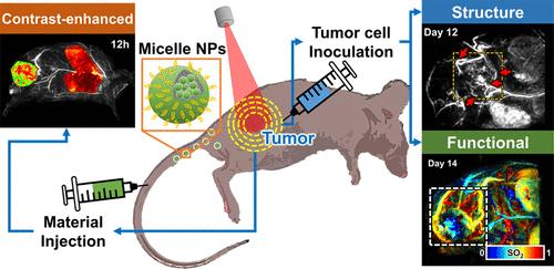
Photoacoustic computed tomography (PACT), an emerging imaging modality in preclinical cancer research, can provide multiparametric 3D information about structures, physiological functions, and pharmacokinetics. Here, we demonstrate the use of high-definition 3D multiparametric PACT imaging of both primary and metastatic tumors in living mice to noninvasively monitor angiogenesis, carcinogenesis, hypoxia, and pharmacokinetics. The high-definition PACT system with a 1024-element hemispherical ultrasound transducer array provides an isotropic spatial resolution of 380 μm, an effective volumetric field-of-view of 12.8 mm × 12.8 mm × 12.8 mm without scanning, and an acquisition time of <30 s for a whole mouse body. Initially, we monitor the structural progression of the tumor microenvironment (e.g., angiogenesis and vessel tortuosity) after tumor cell inoculation. Then, we analyze the change in oxygen saturation of the tumor during carcinogenesis, verifying induced hypoxia in the tumor’s core region. Finally, the whole-body pharmacokinetics are photoacoustically imaged after intravenous injection of micelle-loaded IR780 dye, and the in vivo PACT results are validated in vivo and ex vivo by fluorescence imaging. By employing the premium PACT system and applying multiparametric analyses to subcutaneous primary tumors and metastatic liver tumors, we demonstrate that this PACT system can provide multiparametric analyses for comprehensive cancer research.
期刊介绍:
ACS Nano, published monthly, serves as an international forum for comprehensive articles on nanoscience and nanotechnology research at the intersections of chemistry, biology, materials science, physics, and engineering. The journal fosters communication among scientists in these communities, facilitating collaboration, new research opportunities, and advancements through discoveries. ACS Nano covers synthesis, assembly, characterization, theory, and simulation of nanostructures, nanobiotechnology, nanofabrication, methods and tools for nanoscience and nanotechnology, and self- and directed-assembly. Alongside original research articles, it offers thorough reviews, perspectives on cutting-edge research, and discussions envisioning the future of nanoscience and nanotechnology.


 分享
分享
 求助内容:
求助内容: 应助结果提醒方式:
 扫码关注我们
