{"title":"利用实时采集的 NCCT 图像准确检测和分类颅内脑出血的新型深度学习框架","authors":"Simarjeet Kaur, Amar Singh","doi":"10.1007/s00723-024-01661-z","DOIUrl":null,"url":null,"abstract":"<p>Brain hemorrhage is a critical medical condition that is likely to cause long-term disabilities and death. Timely and precise emergency care, incorporating the accurate interpretation of computed tomography (CT) images, plays a crucial role in the effective management of a hemorrhagic stroke. However, conventional artificial intelligence methods are capable enough to detect the presence or absence of hemorrhage but fail to detect multiple types of hemorrhage with high accuracy. To address this, the paper introduces an innovative Deep Learning based approach that automatically detects, segments, and classifies subtypes of intracranial hemorrhages. The proposed model is trained and evaluated on two different datasets. It is initially trained on a dataset of CT images from the Radiological Society of North America (RSNA) brain CT hemorrhage database, which contained 752,803 head non-contrast computer tomography images obtained from 2,200 patients. Furthermore, the model's performance is validated using a real-time CT dataset collected from a diagnostic lab, comprising 15,000 CT scan images from 176 patients. The proposed model surpasses standard benchmarks for detection and classification, achieving exceptional metrics. It showcases overall segmentation accuracy with a Dice score and Jaccard Index of 0.99 and 0.88 respectively, while the classification metrics include an accuracy of 0.99, precision, recall, and F1 score of 0.97, 0.98, and 0.97 respectively. When two expert radiologists independently assessed the predicted hemorrhage locations and subtypes, ensuring uniform specificity levels, they determined the observed rate of false positives per patient was less. These results validate its applicability as a dependable clinical decision support tool.</p>","PeriodicalId":469,"journal":{"name":"Applied Magnetic Resonance","volume":null,"pages":null},"PeriodicalIF":1.1000,"publicationDate":"2024-06-13","publicationTypes":"Journal Article","fieldsOfStudy":null,"isOpenAccess":false,"openAccessPdf":"","citationCount":"0","resultStr":"{\"title\":\"A New Deep Learning Framework for Accurate Intracranial Brain Hemorrhage Detection and Classification Using Real-Time Collected NCCT Images\",\"authors\":\"Simarjeet Kaur, Amar Singh\",\"doi\":\"10.1007/s00723-024-01661-z\",\"DOIUrl\":null,\"url\":null,\"abstract\":\"<p>Brain hemorrhage is a critical medical condition that is likely to cause long-term disabilities and death. Timely and precise emergency care, incorporating the accurate interpretation of computed tomography (CT) images, plays a crucial role in the effective management of a hemorrhagic stroke. However, conventional artificial intelligence methods are capable enough to detect the presence or absence of hemorrhage but fail to detect multiple types of hemorrhage with high accuracy. To address this, the paper introduces an innovative Deep Learning based approach that automatically detects, segments, and classifies subtypes of intracranial hemorrhages. The proposed model is trained and evaluated on two different datasets. It is initially trained on a dataset of CT images from the Radiological Society of North America (RSNA) brain CT hemorrhage database, which contained 752,803 head non-contrast computer tomography images obtained from 2,200 patients. Furthermore, the model's performance is validated using a real-time CT dataset collected from a diagnostic lab, comprising 15,000 CT scan images from 176 patients. The proposed model surpasses standard benchmarks for detection and classification, achieving exceptional metrics. It showcases overall segmentation accuracy with a Dice score and Jaccard Index of 0.99 and 0.88 respectively, while the classification metrics include an accuracy of 0.99, precision, recall, and F1 score of 0.97, 0.98, and 0.97 respectively. When two expert radiologists independently assessed the predicted hemorrhage locations and subtypes, ensuring uniform specificity levels, they determined the observed rate of false positives per patient was less. These results validate its applicability as a dependable clinical decision support tool.</p>\",\"PeriodicalId\":469,\"journal\":{\"name\":\"Applied Magnetic Resonance\",\"volume\":null,\"pages\":null},\"PeriodicalIF\":1.1000,\"publicationDate\":\"2024-06-13\",\"publicationTypes\":\"Journal Article\",\"fieldsOfStudy\":null,\"isOpenAccess\":false,\"openAccessPdf\":\"\",\"citationCount\":\"0\",\"resultStr\":null,\"platform\":\"Semanticscholar\",\"paperid\":null,\"PeriodicalName\":\"Applied Magnetic Resonance\",\"FirstCategoryId\":\"101\",\"ListUrlMain\":\"https://doi.org/10.1007/s00723-024-01661-z\",\"RegionNum\":4,\"RegionCategory\":\"物理与天体物理\",\"ArticlePicture\":[],\"TitleCN\":null,\"AbstractTextCN\":null,\"PMCID\":null,\"EPubDate\":\"\",\"PubModel\":\"\",\"JCR\":\"Q4\",\"JCRName\":\"PHYSICS, ATOMIC, MOLECULAR & CHEMICAL\",\"Score\":null,\"Total\":0}","platform":"Semanticscholar","paperid":null,"PeriodicalName":"Applied Magnetic Resonance","FirstCategoryId":"101","ListUrlMain":"https://doi.org/10.1007/s00723-024-01661-z","RegionNum":4,"RegionCategory":"物理与天体物理","ArticlePicture":[],"TitleCN":null,"AbstractTextCN":null,"PMCID":null,"EPubDate":"","PubModel":"","JCR":"Q4","JCRName":"PHYSICS, ATOMIC, MOLECULAR & CHEMICAL","Score":null,"Total":0}
A New Deep Learning Framework for Accurate Intracranial Brain Hemorrhage Detection and Classification Using Real-Time Collected NCCT Images
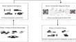
Brain hemorrhage is a critical medical condition that is likely to cause long-term disabilities and death. Timely and precise emergency care, incorporating the accurate interpretation of computed tomography (CT) images, plays a crucial role in the effective management of a hemorrhagic stroke. However, conventional artificial intelligence methods are capable enough to detect the presence or absence of hemorrhage but fail to detect multiple types of hemorrhage with high accuracy. To address this, the paper introduces an innovative Deep Learning based approach that automatically detects, segments, and classifies subtypes of intracranial hemorrhages. The proposed model is trained and evaluated on two different datasets. It is initially trained on a dataset of CT images from the Radiological Society of North America (RSNA) brain CT hemorrhage database, which contained 752,803 head non-contrast computer tomography images obtained from 2,200 patients. Furthermore, the model's performance is validated using a real-time CT dataset collected from a diagnostic lab, comprising 15,000 CT scan images from 176 patients. The proposed model surpasses standard benchmarks for detection and classification, achieving exceptional metrics. It showcases overall segmentation accuracy with a Dice score and Jaccard Index of 0.99 and 0.88 respectively, while the classification metrics include an accuracy of 0.99, precision, recall, and F1 score of 0.97, 0.98, and 0.97 respectively. When two expert radiologists independently assessed the predicted hemorrhage locations and subtypes, ensuring uniform specificity levels, they determined the observed rate of false positives per patient was less. These results validate its applicability as a dependable clinical decision support tool.
期刊介绍:
Applied Magnetic Resonance provides an international forum for the application of magnetic resonance in physics, chemistry, biology, medicine, geochemistry, ecology, engineering, and related fields.
The contents include articles with a strong emphasis on new applications, and on new experimental methods. Additional features include book reviews and Letters to the Editor.


 分享
分享
 求助内容:
求助内容: 应助结果提醒方式:
 扫码关注我们
