John A Carrino, Hamza Ibad, Yenpo Lin, Elena Ghotbi, Joshua Klein, Shadpour Demehri, Filippo Del Grande, Eric Bogner, Mikael P Boesen, Jeffrey H Siewerdsen
{"title":"肌肉骨骼成像中的 CT:仍然有用吗?","authors":"John A Carrino, Hamza Ibad, Yenpo Lin, Elena Ghotbi, Joshua Klein, Shadpour Demehri, Filippo Del Grande, Eric Bogner, Mikael P Boesen, Jeffrey H Siewerdsen","doi":"10.1007/s00256-024-04737-w","DOIUrl":null,"url":null,"abstract":"<p><p>Computed tomography (CT) is a common modality employed for musculoskeletal imaging. Conventional CT techniques are useful for the assessment of trauma in detection, characterization and surgical planning of complex fractures. CT arthrography can depict internal derangement lesions and impact medical decision making of orthopedic providers. In oncology, CT can have a role in the characterization of bone tumors and may elucidate soft tissue mineralization patterns. Several advances in CT technology have led to a variety of acquisition techniques with distinct clinical applications. These include four-dimensional CT, which allows examination of joints during motion; cone-beam CT, which allows examination during physiological weight-bearing conditions; dual-energy CT, which allows material decomposition useful in musculoskeletal deposition disorders (e.g., gout) and bone marrow edema detection; and photon-counting CT, which provides increased spatial resolution, decreased radiation, and material decomposition compared to standard multi-detector CT systems due to its ability to directly translate X-ray photon energies into electrical signals. Advanced acquisition techniques provide higher spatial resolution scans capable of enhanced bony microarchitecture and bone mineral density assessment. Together, these CT acquisition techniques will continue to play a substantial role in the practices of orthopedics, rheumatology, metabolic bone, oncology, and interventional radiology.</p>","PeriodicalId":21783,"journal":{"name":"Skeletal Radiology","volume":" ","pages":"1711-1725"},"PeriodicalIF":2.2000,"publicationDate":"2024-09-01","publicationTypes":"Journal Article","fieldsOfStudy":null,"isOpenAccess":false,"openAccessPdf":"","citationCount":"0","resultStr":"{\"title\":\"CT in musculoskeletal imaging: still helpful and for what?\",\"authors\":\"John A Carrino, Hamza Ibad, Yenpo Lin, Elena Ghotbi, Joshua Klein, Shadpour Demehri, Filippo Del Grande, Eric Bogner, Mikael P Boesen, Jeffrey H Siewerdsen\",\"doi\":\"10.1007/s00256-024-04737-w\",\"DOIUrl\":null,\"url\":null,\"abstract\":\"<p><p>Computed tomography (CT) is a common modality employed for musculoskeletal imaging. Conventional CT techniques are useful for the assessment of trauma in detection, characterization and surgical planning of complex fractures. CT arthrography can depict internal derangement lesions and impact medical decision making of orthopedic providers. In oncology, CT can have a role in the characterization of bone tumors and may elucidate soft tissue mineralization patterns. Several advances in CT technology have led to a variety of acquisition techniques with distinct clinical applications. These include four-dimensional CT, which allows examination of joints during motion; cone-beam CT, which allows examination during physiological weight-bearing conditions; dual-energy CT, which allows material decomposition useful in musculoskeletal deposition disorders (e.g., gout) and bone marrow edema detection; and photon-counting CT, which provides increased spatial resolution, decreased radiation, and material decomposition compared to standard multi-detector CT systems due to its ability to directly translate X-ray photon energies into electrical signals. Advanced acquisition techniques provide higher spatial resolution scans capable of enhanced bony microarchitecture and bone mineral density assessment. Together, these CT acquisition techniques will continue to play a substantial role in the practices of orthopedics, rheumatology, metabolic bone, oncology, and interventional radiology.</p>\",\"PeriodicalId\":21783,\"journal\":{\"name\":\"Skeletal Radiology\",\"volume\":\" \",\"pages\":\"1711-1725\"},\"PeriodicalIF\":2.2000,\"publicationDate\":\"2024-09-01\",\"publicationTypes\":\"Journal Article\",\"fieldsOfStudy\":null,\"isOpenAccess\":false,\"openAccessPdf\":\"\",\"citationCount\":\"0\",\"resultStr\":null,\"platform\":\"Semanticscholar\",\"paperid\":null,\"PeriodicalName\":\"Skeletal Radiology\",\"FirstCategoryId\":\"3\",\"ListUrlMain\":\"https://doi.org/10.1007/s00256-024-04737-w\",\"RegionNum\":3,\"RegionCategory\":\"医学\",\"ArticlePicture\":[],\"TitleCN\":null,\"AbstractTextCN\":null,\"PMCID\":null,\"EPubDate\":\"2024/7/6 0:00:00\",\"PubModel\":\"Epub\",\"JCR\":\"Q2\",\"JCRName\":\"ORTHOPEDICS\",\"Score\":null,\"Total\":0}","platform":"Semanticscholar","paperid":null,"PeriodicalName":"Skeletal Radiology","FirstCategoryId":"3","ListUrlMain":"https://doi.org/10.1007/s00256-024-04737-w","RegionNum":3,"RegionCategory":"医学","ArticlePicture":[],"TitleCN":null,"AbstractTextCN":null,"PMCID":null,"EPubDate":"2024/7/6 0:00:00","PubModel":"Epub","JCR":"Q2","JCRName":"ORTHOPEDICS","Score":null,"Total":0}
CT in musculoskeletal imaging: still helpful and for what?
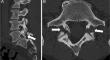
Computed tomography (CT) is a common modality employed for musculoskeletal imaging. Conventional CT techniques are useful for the assessment of trauma in detection, characterization and surgical planning of complex fractures. CT arthrography can depict internal derangement lesions and impact medical decision making of orthopedic providers. In oncology, CT can have a role in the characterization of bone tumors and may elucidate soft tissue mineralization patterns. Several advances in CT technology have led to a variety of acquisition techniques with distinct clinical applications. These include four-dimensional CT, which allows examination of joints during motion; cone-beam CT, which allows examination during physiological weight-bearing conditions; dual-energy CT, which allows material decomposition useful in musculoskeletal deposition disorders (e.g., gout) and bone marrow edema detection; and photon-counting CT, which provides increased spatial resolution, decreased radiation, and material decomposition compared to standard multi-detector CT systems due to its ability to directly translate X-ray photon energies into electrical signals. Advanced acquisition techniques provide higher spatial resolution scans capable of enhanced bony microarchitecture and bone mineral density assessment. Together, these CT acquisition techniques will continue to play a substantial role in the practices of orthopedics, rheumatology, metabolic bone, oncology, and interventional radiology.
期刊介绍:
Skeletal Radiology provides a forum for the dissemination of current knowledge and information dealing with disorders of the musculoskeletal system including the spine. While emphasizing the radiological aspects of the many varied skeletal abnormalities, the journal also adopts an interdisciplinary approach, reflecting the membership of the International Skeletal Society. Thus, the anatomical, pathological, physiological, clinical, metabolic and epidemiological aspects of the many entities affecting the skeleton receive appropriate consideration.
This is the Journal of the International Skeletal Society and the Official Journal of the Society of Skeletal Radiology and the Australasian Musculoskelelal Imaging Group.


 分享
分享
 求助内容:
求助内容: 应助结果提醒方式:
 扫码关注我们
