Sarah Grube, Sarah Latus, Finn Behrendt, Oleksandra Riabova, Maximilian Neidhardt, Alexander Schlaefer
{"title":"利用深度学习在低分辨率超声卷中追踪针头。","authors":"Sarah Grube, Sarah Latus, Finn Behrendt, Oleksandra Riabova, Maximilian Neidhardt, Alexander Schlaefer","doi":"10.1007/s11548-024-03234-8","DOIUrl":null,"url":null,"abstract":"<p><strong>Purpose: </strong>Clinical needle insertion into tissue, commonly assisted by 2D ultrasound imaging for real-time navigation, faces the challenge of precise needle and probe alignment to reduce out-of-plane movement. Recent studies investigate 3D ultrasound imaging together with deep learning to overcome this problem, focusing on acquiring high-resolution images to create optimal conditions for needle tip detection. However, high-resolution also requires a lot of time for image acquisition and processing, which limits the real-time capability. Therefore, we aim to maximize the US volume rate with the trade-off of low image resolution. We propose a deep learning approach to directly extract the 3D needle tip position from sparsely sampled US volumes.</p><p><strong>Methods: </strong>We design an experimental setup with a robot inserting a needle into water and chicken liver tissue. In contrast to manual annotation, we assess the needle tip position from the known robot pose. During insertion, we acquire a large data set of low-resolution volumes using a 16 <math><mo>×</mo></math> 16 element matrix transducer with a volume rate of 4 Hz. We compare the performance of our deep learning approach with conventional needle segmentation.</p><p><strong>Results: </strong>Our experiments in water and liver show that deep learning outperforms the conventional approach while achieving sub-millimeter accuracy. We achieve mean position errors of 0.54 mm in water and 1.54 mm in liver for deep learning.</p><p><strong>Conclusion: </strong>Our study underlines the strengths of deep learning to predict the 3D needle positions from low-resolution ultrasound volumes. This is an important milestone for real-time needle navigation, simplifying the alignment of needle and ultrasound probe and enabling a 3D motion analysis.</p>","PeriodicalId":51251,"journal":{"name":"International Journal of Computer Assisted Radiology and Surgery","volume":" ","pages":"1975-1981"},"PeriodicalIF":2.3000,"publicationDate":"2024-10-01","publicationTypes":"Journal Article","fieldsOfStudy":null,"isOpenAccess":false,"openAccessPdf":"https://www.ncbi.nlm.nih.gov/pmc/articles/PMC11442564/pdf/","citationCount":"0","resultStr":"{\"title\":\"Needle tracking in low-resolution ultrasound volumes using deep learning.\",\"authors\":\"Sarah Grube, Sarah Latus, Finn Behrendt, Oleksandra Riabova, Maximilian Neidhardt, Alexander Schlaefer\",\"doi\":\"10.1007/s11548-024-03234-8\",\"DOIUrl\":null,\"url\":null,\"abstract\":\"<p><strong>Purpose: </strong>Clinical needle insertion into tissue, commonly assisted by 2D ultrasound imaging for real-time navigation, faces the challenge of precise needle and probe alignment to reduce out-of-plane movement. Recent studies investigate 3D ultrasound imaging together with deep learning to overcome this problem, focusing on acquiring high-resolution images to create optimal conditions for needle tip detection. However, high-resolution also requires a lot of time for image acquisition and processing, which limits the real-time capability. Therefore, we aim to maximize the US volume rate with the trade-off of low image resolution. We propose a deep learning approach to directly extract the 3D needle tip position from sparsely sampled US volumes.</p><p><strong>Methods: </strong>We design an experimental setup with a robot inserting a needle into water and chicken liver tissue. In contrast to manual annotation, we assess the needle tip position from the known robot pose. During insertion, we acquire a large data set of low-resolution volumes using a 16 <math><mo>×</mo></math> 16 element matrix transducer with a volume rate of 4 Hz. We compare the performance of our deep learning approach with conventional needle segmentation.</p><p><strong>Results: </strong>Our experiments in water and liver show that deep learning outperforms the conventional approach while achieving sub-millimeter accuracy. We achieve mean position errors of 0.54 mm in water and 1.54 mm in liver for deep learning.</p><p><strong>Conclusion: </strong>Our study underlines the strengths of deep learning to predict the 3D needle positions from low-resolution ultrasound volumes. This is an important milestone for real-time needle navigation, simplifying the alignment of needle and ultrasound probe and enabling a 3D motion analysis.</p>\",\"PeriodicalId\":51251,\"journal\":{\"name\":\"International Journal of Computer Assisted Radiology and Surgery\",\"volume\":\" \",\"pages\":\"1975-1981\"},\"PeriodicalIF\":2.3000,\"publicationDate\":\"2024-10-01\",\"publicationTypes\":\"Journal Article\",\"fieldsOfStudy\":null,\"isOpenAccess\":false,\"openAccessPdf\":\"https://www.ncbi.nlm.nih.gov/pmc/articles/PMC11442564/pdf/\",\"citationCount\":\"0\",\"resultStr\":null,\"platform\":\"Semanticscholar\",\"paperid\":null,\"PeriodicalName\":\"International Journal of Computer Assisted Radiology and Surgery\",\"FirstCategoryId\":\"5\",\"ListUrlMain\":\"https://doi.org/10.1007/s11548-024-03234-8\",\"RegionNum\":3,\"RegionCategory\":\"医学\",\"ArticlePicture\":[],\"TitleCN\":null,\"AbstractTextCN\":null,\"PMCID\":null,\"EPubDate\":\"2024/7/13 0:00:00\",\"PubModel\":\"Epub\",\"JCR\":\"Q3\",\"JCRName\":\"ENGINEERING, BIOMEDICAL\",\"Score\":null,\"Total\":0}","platform":"Semanticscholar","paperid":null,"PeriodicalName":"International Journal of Computer Assisted Radiology and Surgery","FirstCategoryId":"5","ListUrlMain":"https://doi.org/10.1007/s11548-024-03234-8","RegionNum":3,"RegionCategory":"医学","ArticlePicture":[],"TitleCN":null,"AbstractTextCN":null,"PMCID":null,"EPubDate":"2024/7/13 0:00:00","PubModel":"Epub","JCR":"Q3","JCRName":"ENGINEERING, BIOMEDICAL","Score":null,"Total":0}
引用次数: 0
摘要
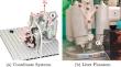
目的:临床上通常通过二维超声成像辅助将针头插入组织进行实时导航,但这面临着针头和探针精确对准以减少平面外移动的挑战。最近的研究将三维超声成像与深度学习相结合来克服这一问题,重点是获取高分辨率图像,为针尖检测创造最佳条件。然而,高分辨率也需要大量时间进行图像采集和处理,从而限制了实时性。因此,我们的目标是在牺牲低图像分辨率的前提下,最大限度地提高 US 容积率。我们提出了一种深度学习方法,从稀疏采样的 US 容量中直接提取三维针尖位置:我们设计了一个实验装置,让机器人将针头插入水和鸡肝组织中。与手动标注不同,我们通过已知的机器人姿势来评估针尖位置。在插入过程中,我们使用 16 × 16 元素矩阵传感器以 4 Hz 的容积率获取了大量低分辨率容积数据集。我们将深度学习方法的性能与传统的针头分割方法进行了比较:结果:我们在水和肝脏中进行的实验表明,深度学习的效果优于传统方法,同时达到了亚毫米级的精度。深度学习在水中的平均位置误差为 0.54 毫米,在肝脏中的平均位置误差为 1.54 毫米:我们的研究强调了深度学习在从低分辨率超声波体积预测三维针位置方面的优势。这是针头实时导航的一个重要里程碑,它简化了针头和超声探头的对准,并实现了三维运动分析。
Needle tracking in low-resolution ultrasound volumes using deep learning.
Purpose: Clinical needle insertion into tissue, commonly assisted by 2D ultrasound imaging for real-time navigation, faces the challenge of precise needle and probe alignment to reduce out-of-plane movement. Recent studies investigate 3D ultrasound imaging together with deep learning to overcome this problem, focusing on acquiring high-resolution images to create optimal conditions for needle tip detection. However, high-resolution also requires a lot of time for image acquisition and processing, which limits the real-time capability. Therefore, we aim to maximize the US volume rate with the trade-off of low image resolution. We propose a deep learning approach to directly extract the 3D needle tip position from sparsely sampled US volumes.
Methods: We design an experimental setup with a robot inserting a needle into water and chicken liver tissue. In contrast to manual annotation, we assess the needle tip position from the known robot pose. During insertion, we acquire a large data set of low-resolution volumes using a 16 16 element matrix transducer with a volume rate of 4 Hz. We compare the performance of our deep learning approach with conventional needle segmentation.
Results: Our experiments in water and liver show that deep learning outperforms the conventional approach while achieving sub-millimeter accuracy. We achieve mean position errors of 0.54 mm in water and 1.54 mm in liver for deep learning.
Conclusion: Our study underlines the strengths of deep learning to predict the 3D needle positions from low-resolution ultrasound volumes. This is an important milestone for real-time needle navigation, simplifying the alignment of needle and ultrasound probe and enabling a 3D motion analysis.
期刊介绍:
The International Journal for Computer Assisted Radiology and Surgery (IJCARS) is a peer-reviewed journal that provides a platform for closing the gap between medical and technical disciplines, and encourages interdisciplinary research and development activities in an international environment.


 分享
分享
 求助内容:
求助内容: 应助结果提醒方式:
 扫码关注我们
