Frederic E Lecouvet, Caroline Chabot, Lokmane Taihi, Thomas Kirchgesner, Perrine Triqueneaux, Jacques Malghem
{"title":"全身核磁共振成像在转移性疾病和骨髓瘤中的现状与未来:如何做以及为什么要做。","authors":"Frederic E Lecouvet, Caroline Chabot, Lokmane Taihi, Thomas Kirchgesner, Perrine Triqueneaux, Jacques Malghem","doi":"10.1007/s00256-024-04723-2","DOIUrl":null,"url":null,"abstract":"<p><p>Metastatic disease and myeloma present unique diagnostic challenges due to their multifocal nature. Accurate detection and staging are critical for determining appropriate treatment. Bone scintigraphy, skeletal radiographs and CT have long been the mainstay for the assessment of these diseases, but have limitations, including reduced sensitivity and radiation exposure. Whole-body MRI has emerged as a highly sensitive and radiation-free alternative imaging modality. Initially developed for skeletal screening, it has extended tumor screening to all organs, providing morphological and physiological information on tumor tissue. Along with PET/CT, whole-body MRI is now accepted for staging and response assessment in many malignancies. It is the first choice in an ever increasing number of cancers (such as myeloma, lobular breast cancer, advanced prostate cancer, myxoid liposarcoma, bone sarcoma, …). It has also been validated as the method of choice for cancer screening in patients with a predisposition to cancer and for staging cancers observed during pregnancy. The current and future challenges for WB-MRI are its availability facing this number of indications, and its acceptance by patients, radiologists and health authorities. Guidelines have been developed to optimize image acquisition and reading, assessment of lesion response to treatment, and to adapt examination designs to specific cancers. The implementation of 3D acquisition, Dixon method, and deep learning-based image optimization further improve the diagnostic performance of the technique and reduce examination durations. Whole-body MRI screening is feasible in less than 30 min. This article reviews validated indications, recent developments, growing acceptance, and future perspectives of whole-body MRI.</p>","PeriodicalId":21783,"journal":{"name":"Skeletal Radiology","volume":" ","pages":"1815-1831"},"PeriodicalIF":2.2000,"publicationDate":"2024-09-01","publicationTypes":"Journal Article","fieldsOfStudy":null,"isOpenAccess":false,"openAccessPdf":"https://www.ncbi.nlm.nih.gov/pmc/articles/PMC11303436/pdf/","citationCount":"0","resultStr":"{\"title\":\"Present and future of whole-body MRI in metastatic disease and myeloma: how and why you will do it.\",\"authors\":\"Frederic E Lecouvet, Caroline Chabot, Lokmane Taihi, Thomas Kirchgesner, Perrine Triqueneaux, Jacques Malghem\",\"doi\":\"10.1007/s00256-024-04723-2\",\"DOIUrl\":null,\"url\":null,\"abstract\":\"<p><p>Metastatic disease and myeloma present unique diagnostic challenges due to their multifocal nature. Accurate detection and staging are critical for determining appropriate treatment. Bone scintigraphy, skeletal radiographs and CT have long been the mainstay for the assessment of these diseases, but have limitations, including reduced sensitivity and radiation exposure. Whole-body MRI has emerged as a highly sensitive and radiation-free alternative imaging modality. Initially developed for skeletal screening, it has extended tumor screening to all organs, providing morphological and physiological information on tumor tissue. Along with PET/CT, whole-body MRI is now accepted for staging and response assessment in many malignancies. It is the first choice in an ever increasing number of cancers (such as myeloma, lobular breast cancer, advanced prostate cancer, myxoid liposarcoma, bone sarcoma, …). It has also been validated as the method of choice for cancer screening in patients with a predisposition to cancer and for staging cancers observed during pregnancy. The current and future challenges for WB-MRI are its availability facing this number of indications, and its acceptance by patients, radiologists and health authorities. Guidelines have been developed to optimize image acquisition and reading, assessment of lesion response to treatment, and to adapt examination designs to specific cancers. The implementation of 3D acquisition, Dixon method, and deep learning-based image optimization further improve the diagnostic performance of the technique and reduce examination durations. Whole-body MRI screening is feasible in less than 30 min. This article reviews validated indications, recent developments, growing acceptance, and future perspectives of whole-body MRI.</p>\",\"PeriodicalId\":21783,\"journal\":{\"name\":\"Skeletal Radiology\",\"volume\":\" \",\"pages\":\"1815-1831\"},\"PeriodicalIF\":2.2000,\"publicationDate\":\"2024-09-01\",\"publicationTypes\":\"Journal Article\",\"fieldsOfStudy\":null,\"isOpenAccess\":false,\"openAccessPdf\":\"https://www.ncbi.nlm.nih.gov/pmc/articles/PMC11303436/pdf/\",\"citationCount\":\"0\",\"resultStr\":null,\"platform\":\"Semanticscholar\",\"paperid\":null,\"PeriodicalName\":\"Skeletal Radiology\",\"FirstCategoryId\":\"3\",\"ListUrlMain\":\"https://doi.org/10.1007/s00256-024-04723-2\",\"RegionNum\":3,\"RegionCategory\":\"医学\",\"ArticlePicture\":[],\"TitleCN\":null,\"AbstractTextCN\":null,\"PMCID\":null,\"EPubDate\":\"2024/7/15 0:00:00\",\"PubModel\":\"Epub\",\"JCR\":\"Q2\",\"JCRName\":\"ORTHOPEDICS\",\"Score\":null,\"Total\":0}","platform":"Semanticscholar","paperid":null,"PeriodicalName":"Skeletal Radiology","FirstCategoryId":"3","ListUrlMain":"https://doi.org/10.1007/s00256-024-04723-2","RegionNum":3,"RegionCategory":"医学","ArticlePicture":[],"TitleCN":null,"AbstractTextCN":null,"PMCID":null,"EPubDate":"2024/7/15 0:00:00","PubModel":"Epub","JCR":"Q2","JCRName":"ORTHOPEDICS","Score":null,"Total":0}
Present and future of whole-body MRI in metastatic disease and myeloma: how and why you will do it.
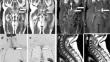
Metastatic disease and myeloma present unique diagnostic challenges due to their multifocal nature. Accurate detection and staging are critical for determining appropriate treatment. Bone scintigraphy, skeletal radiographs and CT have long been the mainstay for the assessment of these diseases, but have limitations, including reduced sensitivity and radiation exposure. Whole-body MRI has emerged as a highly sensitive and radiation-free alternative imaging modality. Initially developed for skeletal screening, it has extended tumor screening to all organs, providing morphological and physiological information on tumor tissue. Along with PET/CT, whole-body MRI is now accepted for staging and response assessment in many malignancies. It is the first choice in an ever increasing number of cancers (such as myeloma, lobular breast cancer, advanced prostate cancer, myxoid liposarcoma, bone sarcoma, …). It has also been validated as the method of choice for cancer screening in patients with a predisposition to cancer and for staging cancers observed during pregnancy. The current and future challenges for WB-MRI are its availability facing this number of indications, and its acceptance by patients, radiologists and health authorities. Guidelines have been developed to optimize image acquisition and reading, assessment of lesion response to treatment, and to adapt examination designs to specific cancers. The implementation of 3D acquisition, Dixon method, and deep learning-based image optimization further improve the diagnostic performance of the technique and reduce examination durations. Whole-body MRI screening is feasible in less than 30 min. This article reviews validated indications, recent developments, growing acceptance, and future perspectives of whole-body MRI.
期刊介绍:
Skeletal Radiology provides a forum for the dissemination of current knowledge and information dealing with disorders of the musculoskeletal system including the spine. While emphasizing the radiological aspects of the many varied skeletal abnormalities, the journal also adopts an interdisciplinary approach, reflecting the membership of the International Skeletal Society. Thus, the anatomical, pathological, physiological, clinical, metabolic and epidemiological aspects of the many entities affecting the skeleton receive appropriate consideration.
This is the Journal of the International Skeletal Society and the Official Journal of the Society of Skeletal Radiology and the Australasian Musculoskelelal Imaging Group.


 分享
分享
 求助内容:
求助内容: 应助结果提醒方式:
 扫码关注我们
