Barbara Hochecker, Katja C Matt, Alica L Meßmer, Melanie M Scherer, Jörg Bergemann
{"title":"利用 Cyto-ID® 染色和细胞成像技术量化人类成纤维细胞中的自噬体","authors":"Barbara Hochecker, Katja C Matt, Alica L Meßmer, Melanie M Scherer, Jörg Bergemann","doi":"10.21769/BioProtoc.5025","DOIUrl":null,"url":null,"abstract":"<p><p><sup>As an essential process for the maintenance of cellular homeostasis and function, autophagy is responsible for the lysosome-mediated degradation of damaged proteins and organelles; therefore, dysregulation of autophagy in humans can lead to a variety of diseases. The link between impaired autophagy and disease highlights the need to investigate possible interventions to address dysregulations. One possible intervention is hyperthermia, which is described in this protocol. To investigate these interventions, a method for absolute quantification of</sup> autophagosomal compartments is required that allows comparison of autophagosomal activity under different conditions. Existing methods such as western blotting and immunohistochemistry for analysing the location and relative abundance of intracellular proteins associated with autophagy, or transmission electron microscopy (TEM), which are either very time-consuming, expensive, or both, are less suitable for this purpose. The method described in this protocol allows the absolute quantification of autophagosomes per cell in human fibroblasts using the CYTO-ID® Autophagy Detection Kit after heat therapy compared to a control. The Cyto-ID® assay is based on the use of a specific dye that selectively stains autophagic compartments, combined with an additional Hoechst 33342 dye for nuclear staining. The subsequent recognition of these stained compartments by the Cytation Imager enables the software to determine the number of autophagosomes per nucleus in living cells. Additionally, this absolute quantification uses an image-based method, and the protocol is easy to use and not time-consuming. Furthermore, the method is not only suitable for heat therapy but can also be adapted to any other desired therapy or substance. Key features • Absolute quantification of autophagic compartments in living cells. • Optimised protocol for the determination of autophagy in primary human skin fibroblasts. • Allows the testing of active substances and treatments concerning autophagy. • Imaging-based method for the determination of autophagy.</p>","PeriodicalId":93907,"journal":{"name":"Bio-protocol","volume":"14 13","pages":"e5025"},"PeriodicalIF":1.3000,"publicationDate":"2024-07-05","publicationTypes":"Journal Article","fieldsOfStudy":null,"isOpenAccess":false,"openAccessPdf":"https://www.ncbi.nlm.nih.gov/pmc/articles/PMC11247374/pdf/","citationCount":"0","resultStr":"{\"title\":\"Quantification of Autophagosomes in Human Fibroblasts Using Cyto-ID<sup>®</sup> Staining and Cytation Imaging.\",\"authors\":\"Barbara Hochecker, Katja C Matt, Alica L Meßmer, Melanie M Scherer, Jörg Bergemann\",\"doi\":\"10.21769/BioProtoc.5025\",\"DOIUrl\":null,\"url\":null,\"abstract\":\"<p><p><sup>As an essential process for the maintenance of cellular homeostasis and function, autophagy is responsible for the lysosome-mediated degradation of damaged proteins and organelles; therefore, dysregulation of autophagy in humans can lead to a variety of diseases. The link between impaired autophagy and disease highlights the need to investigate possible interventions to address dysregulations. One possible intervention is hyperthermia, which is described in this protocol. To investigate these interventions, a method for absolute quantification of</sup> autophagosomal compartments is required that allows comparison of autophagosomal activity under different conditions. Existing methods such as western blotting and immunohistochemistry for analysing the location and relative abundance of intracellular proteins associated with autophagy, or transmission electron microscopy (TEM), which are either very time-consuming, expensive, or both, are less suitable for this purpose. The method described in this protocol allows the absolute quantification of autophagosomes per cell in human fibroblasts using the CYTO-ID® Autophagy Detection Kit after heat therapy compared to a control. The Cyto-ID® assay is based on the use of a specific dye that selectively stains autophagic compartments, combined with an additional Hoechst 33342 dye for nuclear staining. The subsequent recognition of these stained compartments by the Cytation Imager enables the software to determine the number of autophagosomes per nucleus in living cells. Additionally, this absolute quantification uses an image-based method, and the protocol is easy to use and not time-consuming. Furthermore, the method is not only suitable for heat therapy but can also be adapted to any other desired therapy or substance. Key features • Absolute quantification of autophagic compartments in living cells. • Optimised protocol for the determination of autophagy in primary human skin fibroblasts. • Allows the testing of active substances and treatments concerning autophagy. • Imaging-based method for the determination of autophagy.</p>\",\"PeriodicalId\":93907,\"journal\":{\"name\":\"Bio-protocol\",\"volume\":\"14 13\",\"pages\":\"e5025\"},\"PeriodicalIF\":1.3000,\"publicationDate\":\"2024-07-05\",\"publicationTypes\":\"Journal Article\",\"fieldsOfStudy\":null,\"isOpenAccess\":false,\"openAccessPdf\":\"https://www.ncbi.nlm.nih.gov/pmc/articles/PMC11247374/pdf/\",\"citationCount\":\"0\",\"resultStr\":null,\"platform\":\"Semanticscholar\",\"paperid\":null,\"PeriodicalName\":\"Bio-protocol\",\"FirstCategoryId\":\"1085\",\"ListUrlMain\":\"https://doi.org/10.21769/BioProtoc.5025\",\"RegionNum\":0,\"RegionCategory\":null,\"ArticlePicture\":[],\"TitleCN\":null,\"AbstractTextCN\":null,\"PMCID\":null,\"EPubDate\":\"\",\"PubModel\":\"\",\"JCR\":\"Q3\",\"JCRName\":\"BIOLOGY\",\"Score\":null,\"Total\":0}","platform":"Semanticscholar","paperid":null,"PeriodicalName":"Bio-protocol","FirstCategoryId":"1085","ListUrlMain":"https://doi.org/10.21769/BioProtoc.5025","RegionNum":0,"RegionCategory":null,"ArticlePicture":[],"TitleCN":null,"AbstractTextCN":null,"PMCID":null,"EPubDate":"","PubModel":"","JCR":"Q3","JCRName":"BIOLOGY","Score":null,"Total":0}
Quantification of Autophagosomes in Human Fibroblasts Using Cyto-ID® Staining and Cytation Imaging.
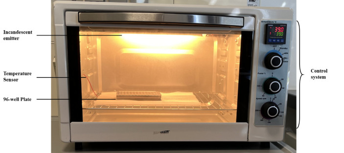
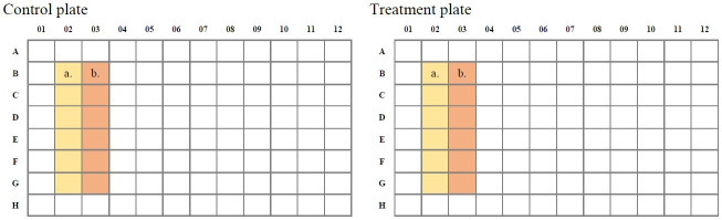
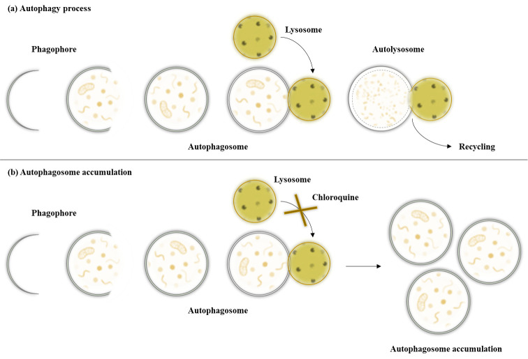
As an essential process for the maintenance of cellular homeostasis and function, autophagy is responsible for the lysosome-mediated degradation of damaged proteins and organelles; therefore, dysregulation of autophagy in humans can lead to a variety of diseases. The link between impaired autophagy and disease highlights the need to investigate possible interventions to address dysregulations. One possible intervention is hyperthermia, which is described in this protocol. To investigate these interventions, a method for absolute quantification of autophagosomal compartments is required that allows comparison of autophagosomal activity under different conditions. Existing methods such as western blotting and immunohistochemistry for analysing the location and relative abundance of intracellular proteins associated with autophagy, or transmission electron microscopy (TEM), which are either very time-consuming, expensive, or both, are less suitable for this purpose. The method described in this protocol allows the absolute quantification of autophagosomes per cell in human fibroblasts using the CYTO-ID® Autophagy Detection Kit after heat therapy compared to a control. The Cyto-ID® assay is based on the use of a specific dye that selectively stains autophagic compartments, combined with an additional Hoechst 33342 dye for nuclear staining. The subsequent recognition of these stained compartments by the Cytation Imager enables the software to determine the number of autophagosomes per nucleus in living cells. Additionally, this absolute quantification uses an image-based method, and the protocol is easy to use and not time-consuming. Furthermore, the method is not only suitable for heat therapy but can also be adapted to any other desired therapy or substance. Key features • Absolute quantification of autophagic compartments in living cells. • Optimised protocol for the determination of autophagy in primary human skin fibroblasts. • Allows the testing of active substances and treatments concerning autophagy. • Imaging-based method for the determination of autophagy.


 分享
分享

 求助内容:
求助内容: 应助结果提醒方式:
 扫码关注我们
