{"title":"磁共振成像在评估肺部恶性肿瘤微波消融术后即时疗效中的价值","authors":"Fandong Zhu, Chen Yang, Jianyun Wang, Tong Zhou, Qianling Li, Subo Wang, Zhenhua Zhao","doi":"10.1097/RTI.0000000000000797","DOIUrl":null,"url":null,"abstract":"<p><strong>Purpose: </strong>To investigate the imaging performance and parametric analysis of magnetic resonance imaging (MRI) immediately after microwave ablation (MWA) of lung malignancies.</p><p><strong>Materials and methods: </strong>We retrospectively analyzed the MRI performance immediately after MWA of 34 cases of lung malignancies. The ablation zone parameters of lung malignancies were measured, including the long diameter (L), short diameter (S), and safety margin of the ablation zone on plain computed tomography (CT), T1-weighted imaging (T1WI), and T2-weighted imaging (T2WI) after MWA. The study calculated the tumor volume (V 0 ), the ablation zone volume (V 1 ), and the ratio of V 0 to V 1 (V%). Statistical differences between the parameters were analyzed.</p><p><strong>Results: </strong>The ablation area of the lesion exhibited central low signal and peripheral high signal on T2WI, central high signal and peripheral equal or high signal on T1WI, and circumferential enhancement in the periphery. The safety margin measured on T2WI was greater than that measured on plain CT and T1WI. On plain CT, the L, S, and V 1 were smaller in the effective treatment group than in the ineffective treatment group ( P <0.05). On T1WI, the V% and safety margin were greater in the effective treatment group than in the ineffective treatment group ( P =0.009 and P =0.016, respectively).</p><p><strong>Conclusions: </strong>MRI may be a new, valuable method to assess immediate efficacy after MWA for lung malignancies using the ablation zone parameters V% on T1WI and safety margin on T2WI.</p>","PeriodicalId":49974,"journal":{"name":"Journal of Thoracic Imaging","volume":" ","pages":"392-398"},"PeriodicalIF":1.3000,"publicationDate":"2024-11-01","publicationTypes":"Journal Article","fieldsOfStudy":null,"isOpenAccess":false,"openAccessPdf":"https://www.ncbi.nlm.nih.gov/pmc/articles/PMC11495527/pdf/","citationCount":"0","resultStr":"{\"title\":\"The Value of Magnetic Resonance Imaging in Assessing Immediate Efficacy After Microwave Ablation of Lung Malignancies.\",\"authors\":\"Fandong Zhu, Chen Yang, Jianyun Wang, Tong Zhou, Qianling Li, Subo Wang, Zhenhua Zhao\",\"doi\":\"10.1097/RTI.0000000000000797\",\"DOIUrl\":null,\"url\":null,\"abstract\":\"<p><strong>Purpose: </strong>To investigate the imaging performance and parametric analysis of magnetic resonance imaging (MRI) immediately after microwave ablation (MWA) of lung malignancies.</p><p><strong>Materials and methods: </strong>We retrospectively analyzed the MRI performance immediately after MWA of 34 cases of lung malignancies. The ablation zone parameters of lung malignancies were measured, including the long diameter (L), short diameter (S), and safety margin of the ablation zone on plain computed tomography (CT), T1-weighted imaging (T1WI), and T2-weighted imaging (T2WI) after MWA. The study calculated the tumor volume (V 0 ), the ablation zone volume (V 1 ), and the ratio of V 0 to V 1 (V%). Statistical differences between the parameters were analyzed.</p><p><strong>Results: </strong>The ablation area of the lesion exhibited central low signal and peripheral high signal on T2WI, central high signal and peripheral equal or high signal on T1WI, and circumferential enhancement in the periphery. The safety margin measured on T2WI was greater than that measured on plain CT and T1WI. On plain CT, the L, S, and V 1 were smaller in the effective treatment group than in the ineffective treatment group ( P <0.05). On T1WI, the V% and safety margin were greater in the effective treatment group than in the ineffective treatment group ( P =0.009 and P =0.016, respectively).</p><p><strong>Conclusions: </strong>MRI may be a new, valuable method to assess immediate efficacy after MWA for lung malignancies using the ablation zone parameters V% on T1WI and safety margin on T2WI.</p>\",\"PeriodicalId\":49974,\"journal\":{\"name\":\"Journal of Thoracic Imaging\",\"volume\":\" \",\"pages\":\"392-398\"},\"PeriodicalIF\":1.3000,\"publicationDate\":\"2024-11-01\",\"publicationTypes\":\"Journal Article\",\"fieldsOfStudy\":null,\"isOpenAccess\":false,\"openAccessPdf\":\"https://www.ncbi.nlm.nih.gov/pmc/articles/PMC11495527/pdf/\",\"citationCount\":\"0\",\"resultStr\":null,\"platform\":\"Semanticscholar\",\"paperid\":null,\"PeriodicalName\":\"Journal of Thoracic Imaging\",\"FirstCategoryId\":\"3\",\"ListUrlMain\":\"https://doi.org/10.1097/RTI.0000000000000797\",\"RegionNum\":4,\"RegionCategory\":\"医学\",\"ArticlePicture\":[],\"TitleCN\":null,\"AbstractTextCN\":null,\"PMCID\":null,\"EPubDate\":\"2024/7/18 0:00:00\",\"PubModel\":\"Epub\",\"JCR\":\"Q3\",\"JCRName\":\"RADIOLOGY, NUCLEAR MEDICINE & MEDICAL IMAGING\",\"Score\":null,\"Total\":0}","platform":"Semanticscholar","paperid":null,"PeriodicalName":"Journal of Thoracic Imaging","FirstCategoryId":"3","ListUrlMain":"https://doi.org/10.1097/RTI.0000000000000797","RegionNum":4,"RegionCategory":"医学","ArticlePicture":[],"TitleCN":null,"AbstractTextCN":null,"PMCID":null,"EPubDate":"2024/7/18 0:00:00","PubModel":"Epub","JCR":"Q3","JCRName":"RADIOLOGY, NUCLEAR MEDICINE & MEDICAL IMAGING","Score":null,"Total":0}
The Value of Magnetic Resonance Imaging in Assessing Immediate Efficacy After Microwave Ablation of Lung Malignancies.
Purpose: To investigate the imaging performance and parametric analysis of magnetic resonance imaging (MRI) immediately after microwave ablation (MWA) of lung malignancies.
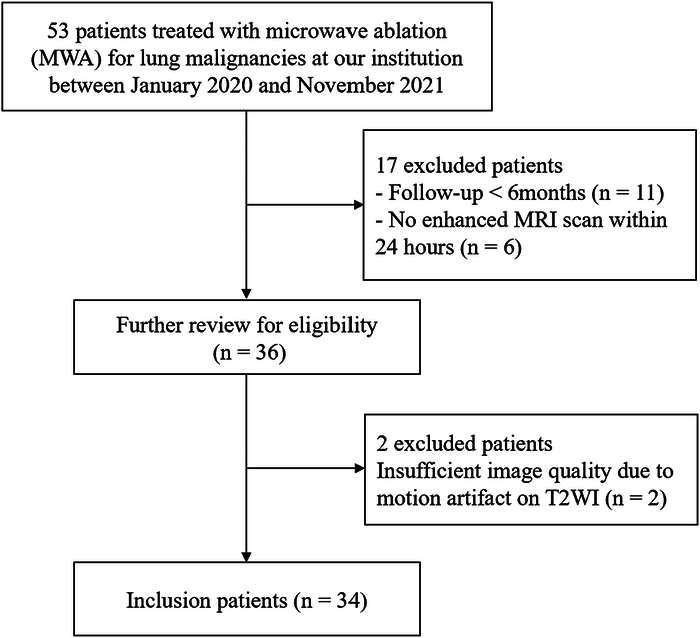
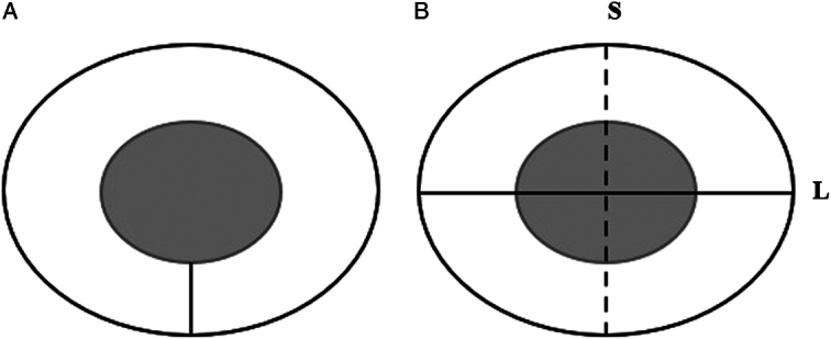
Materials and methods: We retrospectively analyzed the MRI performance immediately after MWA of 34 cases of lung malignancies. The ablation zone parameters of lung malignancies were measured, including the long diameter (L), short diameter (S), and safety margin of the ablation zone on plain computed tomography (CT), T1-weighted imaging (T1WI), and T2-weighted imaging (T2WI) after MWA. The study calculated the tumor volume (V 0 ), the ablation zone volume (V 1 ), and the ratio of V 0 to V 1 (V%). Statistical differences between the parameters were analyzed.
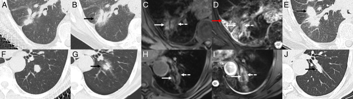
Results: The ablation area of the lesion exhibited central low signal and peripheral high signal on T2WI, central high signal and peripheral equal or high signal on T1WI, and circumferential enhancement in the periphery. The safety margin measured on T2WI was greater than that measured on plain CT and T1WI. On plain CT, the L, S, and V 1 were smaller in the effective treatment group than in the ineffective treatment group ( P <0.05). On T1WI, the V% and safety margin were greater in the effective treatment group than in the ineffective treatment group ( P =0.009 and P =0.016, respectively).
Conclusions: MRI may be a new, valuable method to assess immediate efficacy after MWA for lung malignancies using the ablation zone parameters V% on T1WI and safety margin on T2WI.
期刊介绍:
Journal of Thoracic Imaging (JTI) provides authoritative information on all aspects of the use of imaging techniques in the diagnosis of cardiac and pulmonary diseases. Original articles and analytical reviews published in this timely journal provide the very latest thinking of leading experts concerning the use of chest radiography, computed tomography, magnetic resonance imaging, positron emission tomography, ultrasound, and all other promising imaging techniques in cardiopulmonary radiology.
Official Journal of the Society of Thoracic Radiology:
Japanese Society of Thoracic Radiology
Korean Society of Thoracic Radiology
European Society of Thoracic Imaging.


 分享
分享
 求助内容:
求助内容: 应助结果提醒方式:
 扫码关注我们
