Fernando Duarte, João Neves Silva, Carina Ramos, Colin Hopper
{"title":"正颌手术后咀嚼肌的解剖和功能适应性--3 年随访的核磁共振成像分析。","authors":"Fernando Duarte, João Neves Silva, Carina Ramos, Colin Hopper","doi":"10.1186/s40902-024-00437-6","DOIUrl":null,"url":null,"abstract":"<p><strong>Background: </strong>Orthodontic and surgical technical advances in recent years have resulted in treatment opportunities for a whole range of craniofacial skeletal disorders either in the adolescent or adult patient. In the growing child, these can include myofunctional orthodontic appliance therapy or distraction osteogenesis procedures, while in the adult, the mainstay approach revolves around orthognathic surgery. The literature agrees that for a change in craniofacial morphology to remain stable, the muscles acting upon the facial skeleton must be capable of adaptation in their structure and, therefore, their function. Failure of the muscles to adapt to the change in their length or orientation will place undesirable forces on the muscle attachments leading to potential instability of the skeleton. Adaptation can occur through various processes including those within the neuromuscular feedback mechanism, through changes within muscle structure or through altered muscle physiology, and through changes at the muscle/bone interface. It is now accepted that because there is no single method of assessing masticatory function, several measures should be taken, and whenever possible, simultaneously.</p><p><strong>Methods: </strong>This investigation was designed to apply several, newly developed and more sophisticated methods of measuring muscle structure and function to a situation where adaptation of muscle is pivotal to the success of a therapeutic approach. Patients attending the combined orthodontic/orthognathic surgery clinic at the Clitrofa - Centro Médico, Dentário e Cirúrgico, in Trofa, Portugal, were screened. Ten patients scheduled for a bimaxillary osteotomy involving a combination of maxillary Le Fort I impaction procedure coupled with a sagittal split advancement of the mandible were selected to form the study group. The patients have MRI of the masseter muscle to evaluate the masseter muscle volume and fibre orientation changes. This exam was taken before surgery (T0), 6 to 12 months after surgery (T1), and 3 years after surgery (T2), by two independent observers, according to the protocol jointly developed between the Eastman Dental Institute - University of London and the MRI Centre - Department of Radiology at John Radcliffe Hospital - University of Oxford.</p><p><strong>Results: </strong>Significant differences (p < 0.05) have been identified between Time 0 (pre-op) and Time 1 (6-12 months post-op) regarding the masseter area (mm<sup>2</sup>). The differences against Time 0 (pre-op) seem to disappear at Time 2 (3 years post-op).</p><p><strong>Conclusions: </strong>MRI therefore seems to be a valid tool for measuring differences in the masseter muscle area and volume associated with high-severity occlusal deformities, although showing not to be as efficient in detecting the same differences in cases of low-severity occlusal deformities.</p>","PeriodicalId":18357,"journal":{"name":"Maxillofacial Plastic and Reconstructive Surgery","volume":"46 1","pages":"26"},"PeriodicalIF":2.8000,"publicationDate":"2024-07-19","publicationTypes":"Journal Article","fieldsOfStudy":null,"isOpenAccess":false,"openAccessPdf":"https://www.ncbi.nlm.nih.gov/pmc/articles/PMC11258114/pdf/","citationCount":"0","resultStr":"{\"title\":\"Anatomic and functional masseter muscle adaptation following orthognathic surgery-MRI analysis in 3 years of follow-up.\",\"authors\":\"Fernando Duarte, João Neves Silva, Carina Ramos, Colin Hopper\",\"doi\":\"10.1186/s40902-024-00437-6\",\"DOIUrl\":null,\"url\":null,\"abstract\":\"<p><strong>Background: </strong>Orthodontic and surgical technical advances in recent years have resulted in treatment opportunities for a whole range of craniofacial skeletal disorders either in the adolescent or adult patient. In the growing child, these can include myofunctional orthodontic appliance therapy or distraction osteogenesis procedures, while in the adult, the mainstay approach revolves around orthognathic surgery. The literature agrees that for a change in craniofacial morphology to remain stable, the muscles acting upon the facial skeleton must be capable of adaptation in their structure and, therefore, their function. Failure of the muscles to adapt to the change in their length or orientation will place undesirable forces on the muscle attachments leading to potential instability of the skeleton. Adaptation can occur through various processes including those within the neuromuscular feedback mechanism, through changes within muscle structure or through altered muscle physiology, and through changes at the muscle/bone interface. It is now accepted that because there is no single method of assessing masticatory function, several measures should be taken, and whenever possible, simultaneously.</p><p><strong>Methods: </strong>This investigation was designed to apply several, newly developed and more sophisticated methods of measuring muscle structure and function to a situation where adaptation of muscle is pivotal to the success of a therapeutic approach. Patients attending the combined orthodontic/orthognathic surgery clinic at the Clitrofa - Centro Médico, Dentário e Cirúrgico, in Trofa, Portugal, were screened. Ten patients scheduled for a bimaxillary osteotomy involving a combination of maxillary Le Fort I impaction procedure coupled with a sagittal split advancement of the mandible were selected to form the study group. The patients have MRI of the masseter muscle to evaluate the masseter muscle volume and fibre orientation changes. This exam was taken before surgery (T0), 6 to 12 months after surgery (T1), and 3 years after surgery (T2), by two independent observers, according to the protocol jointly developed between the Eastman Dental Institute - University of London and the MRI Centre - Department of Radiology at John Radcliffe Hospital - University of Oxford.</p><p><strong>Results: </strong>Significant differences (p < 0.05) have been identified between Time 0 (pre-op) and Time 1 (6-12 months post-op) regarding the masseter area (mm<sup>2</sup>). The differences against Time 0 (pre-op) seem to disappear at Time 2 (3 years post-op).</p><p><strong>Conclusions: </strong>MRI therefore seems to be a valid tool for measuring differences in the masseter muscle area and volume associated with high-severity occlusal deformities, although showing not to be as efficient in detecting the same differences in cases of low-severity occlusal deformities.</p>\",\"PeriodicalId\":18357,\"journal\":{\"name\":\"Maxillofacial Plastic and Reconstructive Surgery\",\"volume\":\"46 1\",\"pages\":\"26\"},\"PeriodicalIF\":2.8000,\"publicationDate\":\"2024-07-19\",\"publicationTypes\":\"Journal Article\",\"fieldsOfStudy\":null,\"isOpenAccess\":false,\"openAccessPdf\":\"https://www.ncbi.nlm.nih.gov/pmc/articles/PMC11258114/pdf/\",\"citationCount\":\"0\",\"resultStr\":null,\"platform\":\"Semanticscholar\",\"paperid\":null,\"PeriodicalName\":\"Maxillofacial Plastic and Reconstructive Surgery\",\"FirstCategoryId\":\"1085\",\"ListUrlMain\":\"https://doi.org/10.1186/s40902-024-00437-6\",\"RegionNum\":0,\"RegionCategory\":null,\"ArticlePicture\":[],\"TitleCN\":null,\"AbstractTextCN\":null,\"PMCID\":null,\"EPubDate\":\"\",\"PubModel\":\"\",\"JCR\":\"Q2\",\"JCRName\":\"DENTISTRY, ORAL SURGERY & MEDICINE\",\"Score\":null,\"Total\":0}","platform":"Semanticscholar","paperid":null,"PeriodicalName":"Maxillofacial Plastic and Reconstructive Surgery","FirstCategoryId":"1085","ListUrlMain":"https://doi.org/10.1186/s40902-024-00437-6","RegionNum":0,"RegionCategory":null,"ArticlePicture":[],"TitleCN":null,"AbstractTextCN":null,"PMCID":null,"EPubDate":"","PubModel":"","JCR":"Q2","JCRName":"DENTISTRY, ORAL SURGERY & MEDICINE","Score":null,"Total":0}
引用次数: 0
摘要
背景:近年来,正畸和外科技术的进步为青少年或成人患者治疗各种颅面骨骼疾病提供了机会。对于成长中的儿童,治疗方法包括肌功能正畸矫治器治疗或牵引成骨手术,而对于成人,主要方法是正颌外科手术。文献一致认为,要使颅面形态的变化保持稳定,作用于面部骨骼的肌肉必须能够适应其结构,从而适应其功能。如果肌肉不能适应其长度或方向的变化,就会对肌肉附件产生不良作用力,从而导致骨骼的潜在不稳定性。适应可以通过各种过程发生,包括神经肌肉反馈机制、肌肉结构变化或肌肉生理变化以及肌肉/骨骼界面变化。目前公认的是,由于没有一种评估咀嚼功能的单一方法,因此应采取多种措施,并尽可能同时进行:这项调查旨在将几种新开发的、更复杂的测量肌肉结构和功能的方法应用于肌肉适应性对治疗方法的成功至关重要的情况。在葡萄牙特罗法的 Clitrofa - Centro Médico、Dentário e Cirúrgico 的正畸/正颌外科联合诊所就诊的患者接受了筛查。研究组选取了十名计划接受双颌截骨术的患者,包括上颌勒堡I型咬合植入术和下颌骨矢状劈裂前移术。患者均接受了颌下肌肉核磁共振成像检查,以评估颌下肌肉体积和纤维方向的变化。根据伦敦大学伊士曼牙科研究所和牛津大学约翰-拉德克利夫医院放射科核磁共振成像中心共同制定的方案,由两名独立观察员分别在手术前(T0)、手术后 6 至 12 个月(T1)和手术后 3 年(T2)进行检查:差异显著(P 2)。与时间 0(术前)相比,差异似乎在时间 2(术后 3 年)消失:因此,磁共振成像似乎是测量与高严重性咬合畸形相关的颌下肌面积和体积差异的有效工具,但在检测低严重性咬合畸形病例的相同差异方面,磁共振成像的效率较低。
Anatomic and functional masseter muscle adaptation following orthognathic surgery-MRI analysis in 3 years of follow-up.
Background: Orthodontic and surgical technical advances in recent years have resulted in treatment opportunities for a whole range of craniofacial skeletal disorders either in the adolescent or adult patient. In the growing child, these can include myofunctional orthodontic appliance therapy or distraction osteogenesis procedures, while in the adult, the mainstay approach revolves around orthognathic surgery. The literature agrees that for a change in craniofacial morphology to remain stable, the muscles acting upon the facial skeleton must be capable of adaptation in their structure and, therefore, their function. Failure of the muscles to adapt to the change in their length or orientation will place undesirable forces on the muscle attachments leading to potential instability of the skeleton. Adaptation can occur through various processes including those within the neuromuscular feedback mechanism, through changes within muscle structure or through altered muscle physiology, and through changes at the muscle/bone interface. It is now accepted that because there is no single method of assessing masticatory function, several measures should be taken, and whenever possible, simultaneously.
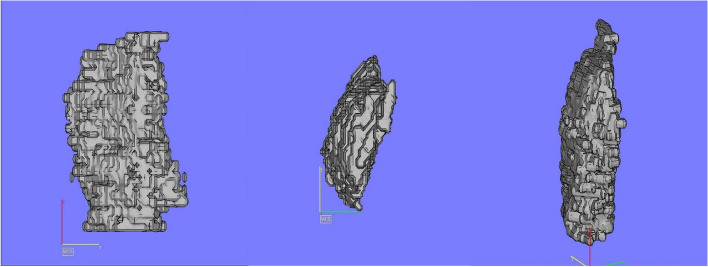
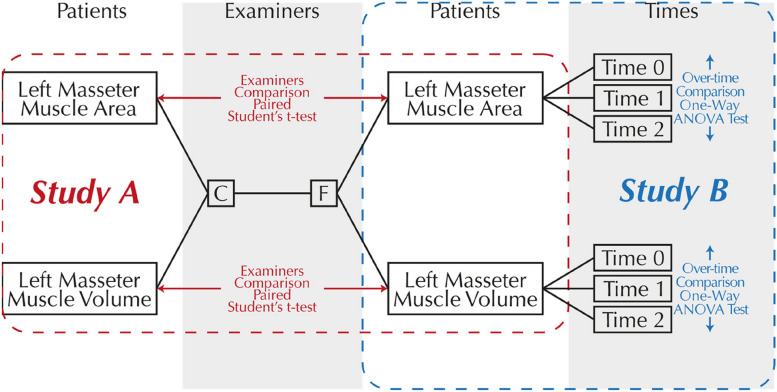
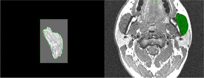
Methods: This investigation was designed to apply several, newly developed and more sophisticated methods of measuring muscle structure and function to a situation where adaptation of muscle is pivotal to the success of a therapeutic approach. Patients attending the combined orthodontic/orthognathic surgery clinic at the Clitrofa - Centro Médico, Dentário e Cirúrgico, in Trofa, Portugal, were screened. Ten patients scheduled for a bimaxillary osteotomy involving a combination of maxillary Le Fort I impaction procedure coupled with a sagittal split advancement of the mandible were selected to form the study group. The patients have MRI of the masseter muscle to evaluate the masseter muscle volume and fibre orientation changes. This exam was taken before surgery (T0), 6 to 12 months after surgery (T1), and 3 years after surgery (T2), by two independent observers, according to the protocol jointly developed between the Eastman Dental Institute - University of London and the MRI Centre - Department of Radiology at John Radcliffe Hospital - University of Oxford.
Results: Significant differences (p < 0.05) have been identified between Time 0 (pre-op) and Time 1 (6-12 months post-op) regarding the masseter area (mm2). The differences against Time 0 (pre-op) seem to disappear at Time 2 (3 years post-op).
Conclusions: MRI therefore seems to be a valid tool for measuring differences in the masseter muscle area and volume associated with high-severity occlusal deformities, although showing not to be as efficient in detecting the same differences in cases of low-severity occlusal deformities.


 分享
分享
 求助内容:
求助内容: 应助结果提醒方式:
 扫码关注我们
