{"title":"生物可吸收磷酸镁水泥椎间融合笼在猪腰椎椎间融合模型中的生物性能:一项可行性研究。","authors":"Xuxuan Wang, Yabin Zhang, Yiguo Wang, Yihao Liu, Xiucan Li, Zhenchuan Han, Yongfei Zhao, Bo Wang, Jianheng Liu, Runsheng Wang, Keya Mao","doi":"10.1007/s00586-024-08387-3","DOIUrl":null,"url":null,"abstract":"<p><strong>Purpose: </strong>The aim of the study was to evaluate the feasibility of a bioabsorbable cage consisting of magnesium and magnesium phosphate cement (MPC) in a porcine lumbar interbody fusion model.</p><p><strong>Methods: </strong>Twelve male Ba-Ma mini pigs underwent lumbar discectomy and fusion with an Mg-MPC cage or a PEEK cage at the L3/L4 and L4/L5 level. Computed tomography (CT) scans were made to evaluate the distractive property by comparing average disc space height (DSH) before and at 6, 12, and 24 weeks after the operation. After the lumbar spines were harvested at 6 or 24 weeks after the operation, micro-CT examination was conducted to analyze the fusion rate, and stiffness of motion segments was investigated through mechanical tests. A histological study was performed to evaluate the tissue type, inflammation, and osteolysis in the intervertebral space.</p><p><strong>Results: </strong>CT scans showed no significant difference between the two groups in average DSH at each time point. Micro-CT scans revealed an equal fusion rate in both groups (0% at 6 weeks, 83.3% at 24 weeks). Both groups showed time-dependent increases in stability, the Mg-MPC cages achieved an inferior stiffness at 6 weeks and a comparable stiffness at 24 weeks. Histologic evaluation showed the presence of newly formed bone in both groups. However, empty spaces were observed at the interface or around the Mg-MPC cages.</p><p><strong>Conclusion: </strong>Compared with the PEEK cages, the Mg-MPC cages achieved comparable distraction, fusion rate, and spinal stability at 24 weeks after the operation. However, due to inferior stiffness at the early stage and fast degradation, further modification of material composition and design are necessary.</p>","PeriodicalId":12323,"journal":{"name":"European Spine Journal","volume":" ","pages":"3324-3333"},"PeriodicalIF":2.7000,"publicationDate":"2024-09-01","publicationTypes":"Journal Article","fieldsOfStudy":null,"isOpenAccess":false,"openAccessPdf":"","citationCount":"0","resultStr":"{\"title\":\"Biological performance of a bioabsorbable magnesium-magnesium phosphate cement interbody fusion cage in a porcine lumbar interbody fusion model: a feasibility study.\",\"authors\":\"Xuxuan Wang, Yabin Zhang, Yiguo Wang, Yihao Liu, Xiucan Li, Zhenchuan Han, Yongfei Zhao, Bo Wang, Jianheng Liu, Runsheng Wang, Keya Mao\",\"doi\":\"10.1007/s00586-024-08387-3\",\"DOIUrl\":null,\"url\":null,\"abstract\":\"<p><strong>Purpose: </strong>The aim of the study was to evaluate the feasibility of a bioabsorbable cage consisting of magnesium and magnesium phosphate cement (MPC) in a porcine lumbar interbody fusion model.</p><p><strong>Methods: </strong>Twelve male Ba-Ma mini pigs underwent lumbar discectomy and fusion with an Mg-MPC cage or a PEEK cage at the L3/L4 and L4/L5 level. Computed tomography (CT) scans were made to evaluate the distractive property by comparing average disc space height (DSH) before and at 6, 12, and 24 weeks after the operation. After the lumbar spines were harvested at 6 or 24 weeks after the operation, micro-CT examination was conducted to analyze the fusion rate, and stiffness of motion segments was investigated through mechanical tests. A histological study was performed to evaluate the tissue type, inflammation, and osteolysis in the intervertebral space.</p><p><strong>Results: </strong>CT scans showed no significant difference between the two groups in average DSH at each time point. Micro-CT scans revealed an equal fusion rate in both groups (0% at 6 weeks, 83.3% at 24 weeks). Both groups showed time-dependent increases in stability, the Mg-MPC cages achieved an inferior stiffness at 6 weeks and a comparable stiffness at 24 weeks. Histologic evaluation showed the presence of newly formed bone in both groups. However, empty spaces were observed at the interface or around the Mg-MPC cages.</p><p><strong>Conclusion: </strong>Compared with the PEEK cages, the Mg-MPC cages achieved comparable distraction, fusion rate, and spinal stability at 24 weeks after the operation. However, due to inferior stiffness at the early stage and fast degradation, further modification of material composition and design are necessary.</p>\",\"PeriodicalId\":12323,\"journal\":{\"name\":\"European Spine Journal\",\"volume\":\" \",\"pages\":\"3324-3333\"},\"PeriodicalIF\":2.7000,\"publicationDate\":\"2024-09-01\",\"publicationTypes\":\"Journal Article\",\"fieldsOfStudy\":null,\"isOpenAccess\":false,\"openAccessPdf\":\"\",\"citationCount\":\"0\",\"resultStr\":null,\"platform\":\"Semanticscholar\",\"paperid\":null,\"PeriodicalName\":\"European Spine Journal\",\"FirstCategoryId\":\"3\",\"ListUrlMain\":\"https://doi.org/10.1007/s00586-024-08387-3\",\"RegionNum\":3,\"RegionCategory\":\"医学\",\"ArticlePicture\":[],\"TitleCN\":null,\"AbstractTextCN\":null,\"PMCID\":null,\"EPubDate\":\"2024/7/22 0:00:00\",\"PubModel\":\"Epub\",\"JCR\":\"Q2\",\"JCRName\":\"CLINICAL NEUROLOGY\",\"Score\":null,\"Total\":0}","platform":"Semanticscholar","paperid":null,"PeriodicalName":"European Spine Journal","FirstCategoryId":"3","ListUrlMain":"https://doi.org/10.1007/s00586-024-08387-3","RegionNum":3,"RegionCategory":"医学","ArticlePicture":[],"TitleCN":null,"AbstractTextCN":null,"PMCID":null,"EPubDate":"2024/7/22 0:00:00","PubModel":"Epub","JCR":"Q2","JCRName":"CLINICAL NEUROLOGY","Score":null,"Total":0}
Biological performance of a bioabsorbable magnesium-magnesium phosphate cement interbody fusion cage in a porcine lumbar interbody fusion model: a feasibility study.
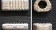
Purpose: The aim of the study was to evaluate the feasibility of a bioabsorbable cage consisting of magnesium and magnesium phosphate cement (MPC) in a porcine lumbar interbody fusion model.
Methods: Twelve male Ba-Ma mini pigs underwent lumbar discectomy and fusion with an Mg-MPC cage or a PEEK cage at the L3/L4 and L4/L5 level. Computed tomography (CT) scans were made to evaluate the distractive property by comparing average disc space height (DSH) before and at 6, 12, and 24 weeks after the operation. After the lumbar spines were harvested at 6 or 24 weeks after the operation, micro-CT examination was conducted to analyze the fusion rate, and stiffness of motion segments was investigated through mechanical tests. A histological study was performed to evaluate the tissue type, inflammation, and osteolysis in the intervertebral space.
Results: CT scans showed no significant difference between the two groups in average DSH at each time point. Micro-CT scans revealed an equal fusion rate in both groups (0% at 6 weeks, 83.3% at 24 weeks). Both groups showed time-dependent increases in stability, the Mg-MPC cages achieved an inferior stiffness at 6 weeks and a comparable stiffness at 24 weeks. Histologic evaluation showed the presence of newly formed bone in both groups. However, empty spaces were observed at the interface or around the Mg-MPC cages.
Conclusion: Compared with the PEEK cages, the Mg-MPC cages achieved comparable distraction, fusion rate, and spinal stability at 24 weeks after the operation. However, due to inferior stiffness at the early stage and fast degradation, further modification of material composition and design are necessary.
期刊介绍:
"European Spine Journal" is a publication founded in response to the increasing trend toward specialization in spinal surgery and spinal pathology in general. The Journal is devoted to all spine related disciplines, including functional and surgical anatomy of the spine, biomechanics and pathophysiology, diagnostic procedures, and neurology, surgery and outcomes. The aim of "European Spine Journal" is to support the further development of highly innovative spine treatments including but not restricted to surgery and to provide an integrated and balanced view of diagnostic, research and treatment procedures as well as outcomes that will enhance effective collaboration among specialists worldwide. The “European Spine Journal” also participates in education by means of videos, interactive meetings and the endorsement of educative efforts.
Official publication of EUROSPINE, The Spine Society of Europe


 分享
分享
 求助内容:
求助内容: 应助结果提醒方式:
 扫码关注我们
