Odair M. Meira, Rolf G. Beutel, Hans Pohl, Thomas van de Kamp, Eduardo A. B. Almeida, Brendon E. Boudinot
{"title":"蜜蜂形态学:Thyreus(膜翅目:猿科)的骨骼肌肉解剖。","authors":"Odair M. Meira, Rolf G. Beutel, Hans Pohl, Thomas van de Kamp, Eduardo A. B. Almeida, Brendon E. Boudinot","doi":"10.1002/jmor.21751","DOIUrl":null,"url":null,"abstract":"<p>Although the knowledge of the skeletal morphology of bees has progressed enormously, a corresponding advance has not happened for the muscular system. Most of the knowledge about bee musculature was generated over 50 years ago, well before the digital revolution for anatomical imaging, including the application of microcomputed tomography. This technique, in particular, has made it possible to dissect small insects digitally, document anatomy efficiently and in detail, and visualize these data three dimensionally. In this study, we document the skeletomuscular system of a cuckoo bee, <i>Thyreus albomaculatus</i> and, with that, we provide a 3D atlas of bee skeletomuscular anatomy. The results obtained for <i>Thyreus</i> are compared with representatives of two other bee families (Andrenidae and Halictidae), to evaluate the generality of our morphological conclusions. Besides documenting 199 specific muscles in terms of origin, insertion, and structure, we update the interpretation of complex homologies in the maxillolabial complex of bee mouthparts. We also clarify the complicated 3D structure of the cephalic endoskeleton, identifying the tentorial, hypostomal, and postgenal structures and their connecting regions. We describe the anatomy of the medial elevator muscles of the head, precisely identifying their origins and insertions as well as their homologs in other groups of Hymenoptera. We reject the hypothesis that the synapomorphic propodeal triangle of Apoidea is homologous with the metapostnotum, and instead recognize that this is a modification of the third phragma. We recognize two previously undocumented metasomal muscle groups in bees, clarifying the serial skeletomusculature of the metasoma and revealing shortcomings of Snodgrass' “internal–external” terminological system for the abdomen. Finally, we elucidate the muscular structure of the sting apparatus, resolving previously unclear interpretations. The work conducted herein not only provides new insights into bee morphology but also represents a source for future phenomic research on Hymenoptera.</p>","PeriodicalId":16528,"journal":{"name":"Journal of Morphology","volume":"285 8","pages":""},"PeriodicalIF":1.4000,"publicationDate":"2024-07-23","publicationTypes":"Journal Article","fieldsOfStudy":null,"isOpenAccess":false,"openAccessPdf":"https://onlinelibrary.wiley.com/doi/epdf/10.1002/jmor.21751","citationCount":"0","resultStr":"{\"title\":\"Bee morphology: A skeletomuscular anatomy of Thyreus (Hymenoptera: Apidae)\",\"authors\":\"Odair M. Meira, Rolf G. Beutel, Hans Pohl, Thomas van de Kamp, Eduardo A. B. Almeida, Brendon E. Boudinot\",\"doi\":\"10.1002/jmor.21751\",\"DOIUrl\":null,\"url\":null,\"abstract\":\"<p>Although the knowledge of the skeletal morphology of bees has progressed enormously, a corresponding advance has not happened for the muscular system. Most of the knowledge about bee musculature was generated over 50 years ago, well before the digital revolution for anatomical imaging, including the application of microcomputed tomography. This technique, in particular, has made it possible to dissect small insects digitally, document anatomy efficiently and in detail, and visualize these data three dimensionally. In this study, we document the skeletomuscular system of a cuckoo bee, <i>Thyreus albomaculatus</i> and, with that, we provide a 3D atlas of bee skeletomuscular anatomy. The results obtained for <i>Thyreus</i> are compared with representatives of two other bee families (Andrenidae and Halictidae), to evaluate the generality of our morphological conclusions. Besides documenting 199 specific muscles in terms of origin, insertion, and structure, we update the interpretation of complex homologies in the maxillolabial complex of bee mouthparts. We also clarify the complicated 3D structure of the cephalic endoskeleton, identifying the tentorial, hypostomal, and postgenal structures and their connecting regions. We describe the anatomy of the medial elevator muscles of the head, precisely identifying their origins and insertions as well as their homologs in other groups of Hymenoptera. We reject the hypothesis that the synapomorphic propodeal triangle of Apoidea is homologous with the metapostnotum, and instead recognize that this is a modification of the third phragma. We recognize two previously undocumented metasomal muscle groups in bees, clarifying the serial skeletomusculature of the metasoma and revealing shortcomings of Snodgrass' “internal–external” terminological system for the abdomen. Finally, we elucidate the muscular structure of the sting apparatus, resolving previously unclear interpretations. The work conducted herein not only provides new insights into bee morphology but also represents a source for future phenomic research on Hymenoptera.</p>\",\"PeriodicalId\":16528,\"journal\":{\"name\":\"Journal of Morphology\",\"volume\":\"285 8\",\"pages\":\"\"},\"PeriodicalIF\":1.4000,\"publicationDate\":\"2024-07-23\",\"publicationTypes\":\"Journal Article\",\"fieldsOfStudy\":null,\"isOpenAccess\":false,\"openAccessPdf\":\"https://onlinelibrary.wiley.com/doi/epdf/10.1002/jmor.21751\",\"citationCount\":\"0\",\"resultStr\":null,\"platform\":\"Semanticscholar\",\"paperid\":null,\"PeriodicalName\":\"Journal of Morphology\",\"FirstCategoryId\":\"3\",\"ListUrlMain\":\"https://onlinelibrary.wiley.com/doi/10.1002/jmor.21751\",\"RegionNum\":4,\"RegionCategory\":\"医学\",\"ArticlePicture\":[],\"TitleCN\":null,\"AbstractTextCN\":null,\"PMCID\":null,\"EPubDate\":\"\",\"PubModel\":\"\",\"JCR\":\"Q2\",\"JCRName\":\"ANATOMY & MORPHOLOGY\",\"Score\":null,\"Total\":0}","platform":"Semanticscholar","paperid":null,"PeriodicalName":"Journal of Morphology","FirstCategoryId":"3","ListUrlMain":"https://onlinelibrary.wiley.com/doi/10.1002/jmor.21751","RegionNum":4,"RegionCategory":"医学","ArticlePicture":[],"TitleCN":null,"AbstractTextCN":null,"PMCID":null,"EPubDate":"","PubModel":"","JCR":"Q2","JCRName":"ANATOMY & MORPHOLOGY","Score":null,"Total":0}
Bee morphology: A skeletomuscular anatomy of Thyreus (Hymenoptera: Apidae)
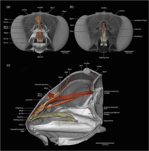
Although the knowledge of the skeletal morphology of bees has progressed enormously, a corresponding advance has not happened for the muscular system. Most of the knowledge about bee musculature was generated over 50 years ago, well before the digital revolution for anatomical imaging, including the application of microcomputed tomography. This technique, in particular, has made it possible to dissect small insects digitally, document anatomy efficiently and in detail, and visualize these data three dimensionally. In this study, we document the skeletomuscular system of a cuckoo bee, Thyreus albomaculatus and, with that, we provide a 3D atlas of bee skeletomuscular anatomy. The results obtained for Thyreus are compared with representatives of two other bee families (Andrenidae and Halictidae), to evaluate the generality of our morphological conclusions. Besides documenting 199 specific muscles in terms of origin, insertion, and structure, we update the interpretation of complex homologies in the maxillolabial complex of bee mouthparts. We also clarify the complicated 3D structure of the cephalic endoskeleton, identifying the tentorial, hypostomal, and postgenal structures and their connecting regions. We describe the anatomy of the medial elevator muscles of the head, precisely identifying their origins and insertions as well as their homologs in other groups of Hymenoptera. We reject the hypothesis that the synapomorphic propodeal triangle of Apoidea is homologous with the metapostnotum, and instead recognize that this is a modification of the third phragma. We recognize two previously undocumented metasomal muscle groups in bees, clarifying the serial skeletomusculature of the metasoma and revealing shortcomings of Snodgrass' “internal–external” terminological system for the abdomen. Finally, we elucidate the muscular structure of the sting apparatus, resolving previously unclear interpretations. The work conducted herein not only provides new insights into bee morphology but also represents a source for future phenomic research on Hymenoptera.
期刊介绍:
The Journal of Morphology welcomes articles of original research in cytology, protozoology, embryology, and general morphology. Articles generally should not exceed 35 printed pages. Preliminary notices or articles of a purely descriptive morphological or taxonomic nature are not included. No paper which has already been published will be accepted, nor will simultaneous publications elsewhere be allowed.
The Journal of Morphology publishes research in functional, comparative, evolutionary and developmental morphology from vertebrates and invertebrates. Human and veterinary anatomy or paleontology are considered when an explicit connection to neontological animal morphology is presented, and the paper contains relevant information for the community of animal morphologists. Based on our long tradition, we continue to seek publishing the best papers in animal morphology.


 分享
分享
 求助内容:
求助内容: 应助结果提醒方式:
 扫码关注我们
