Shawn Shivdat, Tiange Zhan, Alessandro De Palma, Wei-Long Zheng, Parimala Krishnamurthy, Ezhil Paneerselvam, Samuel Snider, Matthew Bevers, Una-May O'Reilly, Jong Woo Lee, M Brandon Westover, Edilberto Amorim
{"title":"心脏骤停后磁共振成像中早期爆发抑制相似性与脑结构损伤严重程度的关系","authors":"Shawn Shivdat, Tiange Zhan, Alessandro De Palma, Wei-Long Zheng, Parimala Krishnamurthy, Ezhil Paneerselvam, Samuel Snider, Matthew Bevers, Una-May O'Reilly, Jong Woo Lee, M Brandon Westover, Edilberto Amorim","doi":"10.1007/s12028-024-02047-6","DOIUrl":null,"url":null,"abstract":"<p><strong>Background: </strong>Identical bursts on electroencephalography (EEG) are considered a specific predictor of poor outcomes in cardiac arrest, but its relationship with structural brain injury severity on magnetic resonance imaging (MRI) is not known.</p><p><strong>Methods: </strong>This was a retrospective analysis of clinical, EEG, and MRI data from adult comatose patients after cardiac arrest. Burst similarity in first 72 h from the time of return of spontaneous circulation were calculated using dynamic time-warping (DTW) for bursts of equal (i.e., 500 ms) and varying (i.e., 100-500 ms) lengths and cross-correlation for bursts of equal lengths. Structural brain injury severity was measured using whole brain mean apparent diffusion coefficient (ADC) on MRI. Pearson's correlation coefficients were calculated between mean burst similarity across consecutive 12-24-h time blocks and mean whole brain ADC values. Good outcome was defined as Cerebral Performance Category of 1-2 (i.e., independence for activities of daily living) at the time of hospital discharge.</p><p><strong>Results: </strong>Of 113 patients with cardiac arrest, 45 patients had burst suppression (mean cardiac arrest to MRI time 4.3 days). Three study participants with burst suppression had a good outcome. Burst similarity calculated using DTW with bursts of varying lengths was correlated with mean ADC value in the first 36 h after cardiac arrest: Pearson's r: 0-12 h: - 0.69 (p = 0.039), 12-24 h: - 0.54 (p = 0.002), 24-36 h: - 0.41 (p = 0.049). Burst similarity measured with bursts of equal lengths was not associated with mean ADC value with cross-correlation or DTW, except for DTW at 60-72 h (- 0.96, p = 0.04).</p><p><strong>Conclusions: </strong>Burst similarity on EEG after cardiac arrest may be associated with acute brain injury severity on MRI. This association was time dependent when measured using DTW.</p>","PeriodicalId":19118,"journal":{"name":"Neurocritical Care","volume":" ","pages":"175-184"},"PeriodicalIF":3.6000,"publicationDate":"2025-02-01","publicationTypes":"Journal Article","fieldsOfStudy":null,"isOpenAccess":false,"openAccessPdf":"https://www.ncbi.nlm.nih.gov/pmc/articles/PMC11757804/pdf/","citationCount":"0","resultStr":"{\"title\":\"Early Burst Suppression Similarity Association with Structural Brain Injury Severity on MRI After Cardiac Arrest.\",\"authors\":\"Shawn Shivdat, Tiange Zhan, Alessandro De Palma, Wei-Long Zheng, Parimala Krishnamurthy, Ezhil Paneerselvam, Samuel Snider, Matthew Bevers, Una-May O'Reilly, Jong Woo Lee, M Brandon Westover, Edilberto Amorim\",\"doi\":\"10.1007/s12028-024-02047-6\",\"DOIUrl\":null,\"url\":null,\"abstract\":\"<p><strong>Background: </strong>Identical bursts on electroencephalography (EEG) are considered a specific predictor of poor outcomes in cardiac arrest, but its relationship with structural brain injury severity on magnetic resonance imaging (MRI) is not known.</p><p><strong>Methods: </strong>This was a retrospective analysis of clinical, EEG, and MRI data from adult comatose patients after cardiac arrest. Burst similarity in first 72 h from the time of return of spontaneous circulation were calculated using dynamic time-warping (DTW) for bursts of equal (i.e., 500 ms) and varying (i.e., 100-500 ms) lengths and cross-correlation for bursts of equal lengths. Structural brain injury severity was measured using whole brain mean apparent diffusion coefficient (ADC) on MRI. Pearson's correlation coefficients were calculated between mean burst similarity across consecutive 12-24-h time blocks and mean whole brain ADC values. Good outcome was defined as Cerebral Performance Category of 1-2 (i.e., independence for activities of daily living) at the time of hospital discharge.</p><p><strong>Results: </strong>Of 113 patients with cardiac arrest, 45 patients had burst suppression (mean cardiac arrest to MRI time 4.3 days). Three study participants with burst suppression had a good outcome. Burst similarity calculated using DTW with bursts of varying lengths was correlated with mean ADC value in the first 36 h after cardiac arrest: Pearson's r: 0-12 h: - 0.69 (p = 0.039), 12-24 h: - 0.54 (p = 0.002), 24-36 h: - 0.41 (p = 0.049). Burst similarity measured with bursts of equal lengths was not associated with mean ADC value with cross-correlation or DTW, except for DTW at 60-72 h (- 0.96, p = 0.04).</p><p><strong>Conclusions: </strong>Burst similarity on EEG after cardiac arrest may be associated with acute brain injury severity on MRI. This association was time dependent when measured using DTW.</p>\",\"PeriodicalId\":19118,\"journal\":{\"name\":\"Neurocritical Care\",\"volume\":\" \",\"pages\":\"175-184\"},\"PeriodicalIF\":3.6000,\"publicationDate\":\"2025-02-01\",\"publicationTypes\":\"Journal Article\",\"fieldsOfStudy\":null,\"isOpenAccess\":false,\"openAccessPdf\":\"https://www.ncbi.nlm.nih.gov/pmc/articles/PMC11757804/pdf/\",\"citationCount\":\"0\",\"resultStr\":null,\"platform\":\"Semanticscholar\",\"paperid\":null,\"PeriodicalName\":\"Neurocritical Care\",\"FirstCategoryId\":\"3\",\"ListUrlMain\":\"https://doi.org/10.1007/s12028-024-02047-6\",\"RegionNum\":3,\"RegionCategory\":\"医学\",\"ArticlePicture\":[],\"TitleCN\":null,\"AbstractTextCN\":null,\"PMCID\":null,\"EPubDate\":\"2024/7/24 0:00:00\",\"PubModel\":\"Epub\",\"JCR\":\"Q2\",\"JCRName\":\"CLINICAL NEUROLOGY\",\"Score\":null,\"Total\":0}","platform":"Semanticscholar","paperid":null,"PeriodicalName":"Neurocritical Care","FirstCategoryId":"3","ListUrlMain":"https://doi.org/10.1007/s12028-024-02047-6","RegionNum":3,"RegionCategory":"医学","ArticlePicture":[],"TitleCN":null,"AbstractTextCN":null,"PMCID":null,"EPubDate":"2024/7/24 0:00:00","PubModel":"Epub","JCR":"Q2","JCRName":"CLINICAL NEUROLOGY","Score":null,"Total":0}
Early Burst Suppression Similarity Association with Structural Brain Injury Severity on MRI After Cardiac Arrest.
Background: Identical bursts on electroencephalography (EEG) are considered a specific predictor of poor outcomes in cardiac arrest, but its relationship with structural brain injury severity on magnetic resonance imaging (MRI) is not known.
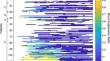
Methods: This was a retrospective analysis of clinical, EEG, and MRI data from adult comatose patients after cardiac arrest. Burst similarity in first 72 h from the time of return of spontaneous circulation were calculated using dynamic time-warping (DTW) for bursts of equal (i.e., 500 ms) and varying (i.e., 100-500 ms) lengths and cross-correlation for bursts of equal lengths. Structural brain injury severity was measured using whole brain mean apparent diffusion coefficient (ADC) on MRI. Pearson's correlation coefficients were calculated between mean burst similarity across consecutive 12-24-h time blocks and mean whole brain ADC values. Good outcome was defined as Cerebral Performance Category of 1-2 (i.e., independence for activities of daily living) at the time of hospital discharge.
Results: Of 113 patients with cardiac arrest, 45 patients had burst suppression (mean cardiac arrest to MRI time 4.3 days). Three study participants with burst suppression had a good outcome. Burst similarity calculated using DTW with bursts of varying lengths was correlated with mean ADC value in the first 36 h after cardiac arrest: Pearson's r: 0-12 h: - 0.69 (p = 0.039), 12-24 h: - 0.54 (p = 0.002), 24-36 h: - 0.41 (p = 0.049). Burst similarity measured with bursts of equal lengths was not associated with mean ADC value with cross-correlation or DTW, except for DTW at 60-72 h (- 0.96, p = 0.04).
Conclusions: Burst similarity on EEG after cardiac arrest may be associated with acute brain injury severity on MRI. This association was time dependent when measured using DTW.
期刊介绍:
Neurocritical Care is a peer reviewed scientific publication whose major goal is to disseminate new knowledge on all aspects of acute neurological care. It is directed towards neurosurgeons, neuro-intensivists, neurologists, anesthesiologists, emergency physicians, and critical care nurses treating patients with urgent neurologic disorders. These are conditions that may potentially evolve rapidly and could need immediate medical or surgical intervention. Neurocritical Care provides a comprehensive overview of current developments in intensive care neurology, neurosurgery and neuroanesthesia and includes information about new therapeutic avenues and technological innovations. Neurocritical Care is the official journal of the Neurocritical Care Society.


 分享
分享
 求助内容:
求助内容: 应助结果提醒方式:
 扫码关注我们
