{"title":"以磁共振成像为基础的机器学习驱动脑年龄模型,用于对轻度认知障碍转换者进行分类。","authors":"Hanna Lu, Jing Li","doi":"10.1177/11795735241266556","DOIUrl":null,"url":null,"abstract":"<p><strong>Background: </strong>Brain age model, including estimated brain age and brain-predicted age difference (brain-PAD), has shown great potentials for serving as imaging markers for monitoring normal ageing, as well as for identifying the individuals in the pre-diagnostic phase of neurodegenerative diseases.</p><p><strong>Purpose: </strong>This study aimed to investigate the brain age models in normal ageing and mild cognitive impairments (MCI) converters and their values in classifying MCI conversion.</p><p><strong>Methods: </strong>Pre-trained brain age model was constructed using the structural magnetic resonance imaging (MRI) data from the Cambridge Centre for Ageing and Neuroscience (Cam-CAN) project (N = 609). The tested brain age model was built using the baseline, 1-year and 3-year follow-up MRI data from normal ageing (NA) adults (n = 32) and MCI converters (n = 22) drew from the Open Access Series of Imaging Studies (OASIS-2). The quantitative measures of morphometry included total intracranial volume (TIV), gray matter volume (GMV) and cortical thickness. Brain age models were calculated based on the individual's morphometric features using the support vector machine (SVM) algorithm.</p><p><strong>Results: </strong>With comparable chronological age, MCI converters showed significant increased TIV-based (Baseline: <i>P</i> = 0.021; 1-year follow-up: <i>P</i> = 0.037; 3-year follow-up: <i>P</i> = 0.001) and left GMV-based brain age than NA adults at all time points. Higher brain-PAD scores were associated with worse global cognition. Acceptable classification performance of TIV-based (AUC = 0.698) and left GMV-based brain age (AUC = 0.703) was found, which could differentiate the MCI converters from NA adults at the baseline.</p><p><strong>Conclusions: </strong>This is the first demonstration that MRI-informed brain age models exhibit feature-specific patterns. The greater GMV-based brain age observed in MCI converters may provide new evidence for identifying the individuals at the early stage of neurodegeneration. Our findings added value to existing quantitative imaging markers and might help to improve disease monitoring and accelerate personalized treatments in clinical practice.</p>","PeriodicalId":15218,"journal":{"name":"Journal of Central Nervous System Disease","volume":"16 ","pages":"11795735241266556"},"PeriodicalIF":2.8000,"publicationDate":"2024-07-21","publicationTypes":"Journal Article","fieldsOfStudy":null,"isOpenAccess":false,"openAccessPdf":"https://www.ncbi.nlm.nih.gov/pmc/articles/PMC11268046/pdf/","citationCount":"0","resultStr":"{\"title\":\"MRI-informed machine learning-driven brain age models for classifying mild cognitive impairment converters.\",\"authors\":\"Hanna Lu, Jing Li\",\"doi\":\"10.1177/11795735241266556\",\"DOIUrl\":null,\"url\":null,\"abstract\":\"<p><strong>Background: </strong>Brain age model, including estimated brain age and brain-predicted age difference (brain-PAD), has shown great potentials for serving as imaging markers for monitoring normal ageing, as well as for identifying the individuals in the pre-diagnostic phase of neurodegenerative diseases.</p><p><strong>Purpose: </strong>This study aimed to investigate the brain age models in normal ageing and mild cognitive impairments (MCI) converters and their values in classifying MCI conversion.</p><p><strong>Methods: </strong>Pre-trained brain age model was constructed using the structural magnetic resonance imaging (MRI) data from the Cambridge Centre for Ageing and Neuroscience (Cam-CAN) project (N = 609). The tested brain age model was built using the baseline, 1-year and 3-year follow-up MRI data from normal ageing (NA) adults (n = 32) and MCI converters (n = 22) drew from the Open Access Series of Imaging Studies (OASIS-2). The quantitative measures of morphometry included total intracranial volume (TIV), gray matter volume (GMV) and cortical thickness. Brain age models were calculated based on the individual's morphometric features using the support vector machine (SVM) algorithm.</p><p><strong>Results: </strong>With comparable chronological age, MCI converters showed significant increased TIV-based (Baseline: <i>P</i> = 0.021; 1-year follow-up: <i>P</i> = 0.037; 3-year follow-up: <i>P</i> = 0.001) and left GMV-based brain age than NA adults at all time points. Higher brain-PAD scores were associated with worse global cognition. Acceptable classification performance of TIV-based (AUC = 0.698) and left GMV-based brain age (AUC = 0.703) was found, which could differentiate the MCI converters from NA adults at the baseline.</p><p><strong>Conclusions: </strong>This is the first demonstration that MRI-informed brain age models exhibit feature-specific patterns. The greater GMV-based brain age observed in MCI converters may provide new evidence for identifying the individuals at the early stage of neurodegeneration. Our findings added value to existing quantitative imaging markers and might help to improve disease monitoring and accelerate personalized treatments in clinical practice.</p>\",\"PeriodicalId\":15218,\"journal\":{\"name\":\"Journal of Central Nervous System Disease\",\"volume\":\"16 \",\"pages\":\"11795735241266556\"},\"PeriodicalIF\":2.8000,\"publicationDate\":\"2024-07-21\",\"publicationTypes\":\"Journal Article\",\"fieldsOfStudy\":null,\"isOpenAccess\":false,\"openAccessPdf\":\"https://www.ncbi.nlm.nih.gov/pmc/articles/PMC11268046/pdf/\",\"citationCount\":\"0\",\"resultStr\":null,\"platform\":\"Semanticscholar\",\"paperid\":null,\"PeriodicalName\":\"Journal of Central Nervous System Disease\",\"FirstCategoryId\":\"1085\",\"ListUrlMain\":\"https://doi.org/10.1177/11795735241266556\",\"RegionNum\":0,\"RegionCategory\":null,\"ArticlePicture\":[],\"TitleCN\":null,\"AbstractTextCN\":null,\"PMCID\":null,\"EPubDate\":\"2024/1/1 0:00:00\",\"PubModel\":\"eCollection\",\"JCR\":\"Q2\",\"JCRName\":\"CLINICAL NEUROLOGY\",\"Score\":null,\"Total\":0}","platform":"Semanticscholar","paperid":null,"PeriodicalName":"Journal of Central Nervous System Disease","FirstCategoryId":"1085","ListUrlMain":"https://doi.org/10.1177/11795735241266556","RegionNum":0,"RegionCategory":null,"ArticlePicture":[],"TitleCN":null,"AbstractTextCN":null,"PMCID":null,"EPubDate":"2024/1/1 0:00:00","PubModel":"eCollection","JCR":"Q2","JCRName":"CLINICAL NEUROLOGY","Score":null,"Total":0}
MRI-informed machine learning-driven brain age models for classifying mild cognitive impairment converters.
Background: Brain age model, including estimated brain age and brain-predicted age difference (brain-PAD), has shown great potentials for serving as imaging markers for monitoring normal ageing, as well as for identifying the individuals in the pre-diagnostic phase of neurodegenerative diseases.
Purpose: This study aimed to investigate the brain age models in normal ageing and mild cognitive impairments (MCI) converters and their values in classifying MCI conversion.
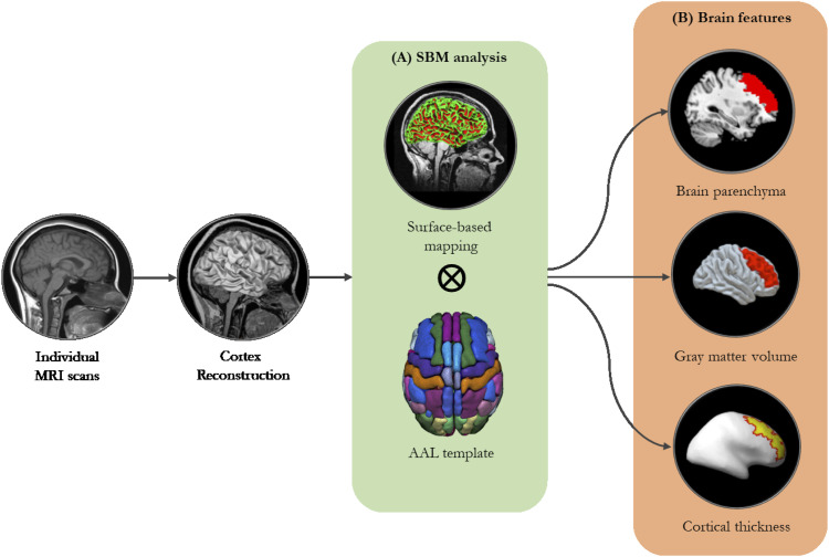
Methods: Pre-trained brain age model was constructed using the structural magnetic resonance imaging (MRI) data from the Cambridge Centre for Ageing and Neuroscience (Cam-CAN) project (N = 609). The tested brain age model was built using the baseline, 1-year and 3-year follow-up MRI data from normal ageing (NA) adults (n = 32) and MCI converters (n = 22) drew from the Open Access Series of Imaging Studies (OASIS-2). The quantitative measures of morphometry included total intracranial volume (TIV), gray matter volume (GMV) and cortical thickness. Brain age models were calculated based on the individual's morphometric features using the support vector machine (SVM) algorithm.
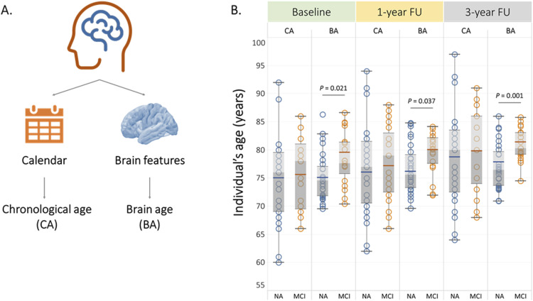
Results: With comparable chronological age, MCI converters showed significant increased TIV-based (Baseline: P = 0.021; 1-year follow-up: P = 0.037; 3-year follow-up: P = 0.001) and left GMV-based brain age than NA adults at all time points. Higher brain-PAD scores were associated with worse global cognition. Acceptable classification performance of TIV-based (AUC = 0.698) and left GMV-based brain age (AUC = 0.703) was found, which could differentiate the MCI converters from NA adults at the baseline.
Conclusions: This is the first demonstration that MRI-informed brain age models exhibit feature-specific patterns. The greater GMV-based brain age observed in MCI converters may provide new evidence for identifying the individuals at the early stage of neurodegeneration. Our findings added value to existing quantitative imaging markers and might help to improve disease monitoring and accelerate personalized treatments in clinical practice.


 分享
分享
 求助内容:
求助内容: 应助结果提醒方式:
 扫码关注我们
