Anne Cathrine Scherer-Quenzer, Inga Beyers, Adam Kalisz, Stephanie Tina Sauer, Marcus Zimmermann, Achim Wöckel, Bülent Polat, Tanja Schlaiss, Selina Schelbert, Matthias Kiesel
{"title":"评估个性化 3D 打印模型在宫颈癌检查、诊断和治疗规划中的价值。","authors":"Anne Cathrine Scherer-Quenzer, Inga Beyers, Adam Kalisz, Stephanie Tina Sauer, Marcus Zimmermann, Achim Wöckel, Bülent Polat, Tanja Schlaiss, Selina Schelbert, Matthias Kiesel","doi":"10.1186/s41205-024-00229-8","DOIUrl":null,"url":null,"abstract":"<p><strong>Background: </strong>3D printing holds great potential of improving examination, diagnosis and treatment planning as well as interprofessional communication in the field of gynecological oncology. In the current manuscript we evaluated five individualized, patient-specific models of cervical cancer FIGO Stage I-III, created with 3D printing, concerning their value for translational oncology.</p><p><strong>Methods: </strong>Magnetic resonance imaging (MRI) of the pelvis was performed on a 3.0 Tesla MRI, including a T2-weighted isotropic 3D sequence. The MRI images were segmented and transferred to virtual 3D models via a custom-built 3D-model generation pipeline and printed by material extrusion. The 3D models were evaluated by all medical specialties involved in patient care of cervical cancer, namely surgeons, radiologists, pathologists and radiation oncologists. Information was obtained from evaluated profession-specific questionnaires which were filled out after inspecting all five models. The questionnaires included multiple-select questions, questions based on Likert scales (1 = \"strongly disagree \" or \"not at all useful \" up to 5 = \"strongly agree \" or \"extremely useful \") and dichotomous questions (\"Yes\" or \"No\").</p><p><strong>Results: </strong>Surgeons rated the models as useful during surgery (4.0 out of 5) and for patient communication (4.7 out of 5). Furthermore, they believed that the models had the potential to revise the patients' treatment plan (3.7 out of 5). Pathologists evaluated with mean ratings of 3.0 out of 5 for the usefulness of the models in diagnostic reporting and macroscopic evaluation. Radiologist acknowledged the possibility of providing additional information compared to imaging alone (3.7 out of 5). Radiation oncologists strongly supported the concept by rating the models highly for understanding patient-specific pathological characteristics (4.3 out of 5), assisting interprofessional communication (mean 4.3 out of 5) and communication with patients (4.7 out of 5). They also found the models useful for improving radiotherapy treatment planning (4.3 out of 5).</p><p><strong>Conclusion: </strong>The study revealed that the 3D printed models were generally well-received by all medical disciplines, with radiation oncologists showing particularly strong support. Addressing the concerns and tailoring the use of 3D models to the specific needs of each medical speciality will be essential for realizing their full potential in clinical practice.</p>","PeriodicalId":72036,"journal":{"name":"3D printing in medicine","volume":"10 1","pages":"25"},"PeriodicalIF":3.1000,"publicationDate":"2024-07-27","publicationTypes":"Journal Article","fieldsOfStudy":null,"isOpenAccess":false,"openAccessPdf":"https://www.ncbi.nlm.nih.gov/pmc/articles/PMC11282658/pdf/","citationCount":"0","resultStr":"{\"title\":\"Evaluating the value of individualized 3D printed models for examination, diagnosis and treatment planning of cervical cancer.\",\"authors\":\"Anne Cathrine Scherer-Quenzer, Inga Beyers, Adam Kalisz, Stephanie Tina Sauer, Marcus Zimmermann, Achim Wöckel, Bülent Polat, Tanja Schlaiss, Selina Schelbert, Matthias Kiesel\",\"doi\":\"10.1186/s41205-024-00229-8\",\"DOIUrl\":null,\"url\":null,\"abstract\":\"<p><strong>Background: </strong>3D printing holds great potential of improving examination, diagnosis and treatment planning as well as interprofessional communication in the field of gynecological oncology. In the current manuscript we evaluated five individualized, patient-specific models of cervical cancer FIGO Stage I-III, created with 3D printing, concerning their value for translational oncology.</p><p><strong>Methods: </strong>Magnetic resonance imaging (MRI) of the pelvis was performed on a 3.0 Tesla MRI, including a T2-weighted isotropic 3D sequence. The MRI images were segmented and transferred to virtual 3D models via a custom-built 3D-model generation pipeline and printed by material extrusion. The 3D models were evaluated by all medical specialties involved in patient care of cervical cancer, namely surgeons, radiologists, pathologists and radiation oncologists. Information was obtained from evaluated profession-specific questionnaires which were filled out after inspecting all five models. The questionnaires included multiple-select questions, questions based on Likert scales (1 = \\\"strongly disagree \\\" or \\\"not at all useful \\\" up to 5 = \\\"strongly agree \\\" or \\\"extremely useful \\\") and dichotomous questions (\\\"Yes\\\" or \\\"No\\\").</p><p><strong>Results: </strong>Surgeons rated the models as useful during surgery (4.0 out of 5) and for patient communication (4.7 out of 5). Furthermore, they believed that the models had the potential to revise the patients' treatment plan (3.7 out of 5). Pathologists evaluated with mean ratings of 3.0 out of 5 for the usefulness of the models in diagnostic reporting and macroscopic evaluation. Radiologist acknowledged the possibility of providing additional information compared to imaging alone (3.7 out of 5). Radiation oncologists strongly supported the concept by rating the models highly for understanding patient-specific pathological characteristics (4.3 out of 5), assisting interprofessional communication (mean 4.3 out of 5) and communication with patients (4.7 out of 5). They also found the models useful for improving radiotherapy treatment planning (4.3 out of 5).</p><p><strong>Conclusion: </strong>The study revealed that the 3D printed models were generally well-received by all medical disciplines, with radiation oncologists showing particularly strong support. Addressing the concerns and tailoring the use of 3D models to the specific needs of each medical speciality will be essential for realizing their full potential in clinical practice.</p>\",\"PeriodicalId\":72036,\"journal\":{\"name\":\"3D printing in medicine\",\"volume\":\"10 1\",\"pages\":\"25\"},\"PeriodicalIF\":3.1000,\"publicationDate\":\"2024-07-27\",\"publicationTypes\":\"Journal Article\",\"fieldsOfStudy\":null,\"isOpenAccess\":false,\"openAccessPdf\":\"https://www.ncbi.nlm.nih.gov/pmc/articles/PMC11282658/pdf/\",\"citationCount\":\"0\",\"resultStr\":null,\"platform\":\"Semanticscholar\",\"paperid\":null,\"PeriodicalName\":\"3D printing in medicine\",\"FirstCategoryId\":\"1085\",\"ListUrlMain\":\"https://doi.org/10.1186/s41205-024-00229-8\",\"RegionNum\":0,\"RegionCategory\":null,\"ArticlePicture\":[],\"TitleCN\":null,\"AbstractTextCN\":null,\"PMCID\":null,\"EPubDate\":\"\",\"PubModel\":\"\",\"JCR\":\"Q1\",\"JCRName\":\"RADIOLOGY, NUCLEAR MEDICINE & MEDICAL IMAGING\",\"Score\":null,\"Total\":0}","platform":"Semanticscholar","paperid":null,"PeriodicalName":"3D printing in medicine","FirstCategoryId":"1085","ListUrlMain":"https://doi.org/10.1186/s41205-024-00229-8","RegionNum":0,"RegionCategory":null,"ArticlePicture":[],"TitleCN":null,"AbstractTextCN":null,"PMCID":null,"EPubDate":"","PubModel":"","JCR":"Q1","JCRName":"RADIOLOGY, NUCLEAR MEDICINE & MEDICAL IMAGING","Score":null,"Total":0}
Evaluating the value of individualized 3D printed models for examination, diagnosis and treatment planning of cervical cancer.
Background: 3D printing holds great potential of improving examination, diagnosis and treatment planning as well as interprofessional communication in the field of gynecological oncology. In the current manuscript we evaluated five individualized, patient-specific models of cervical cancer FIGO Stage I-III, created with 3D printing, concerning their value for translational oncology.
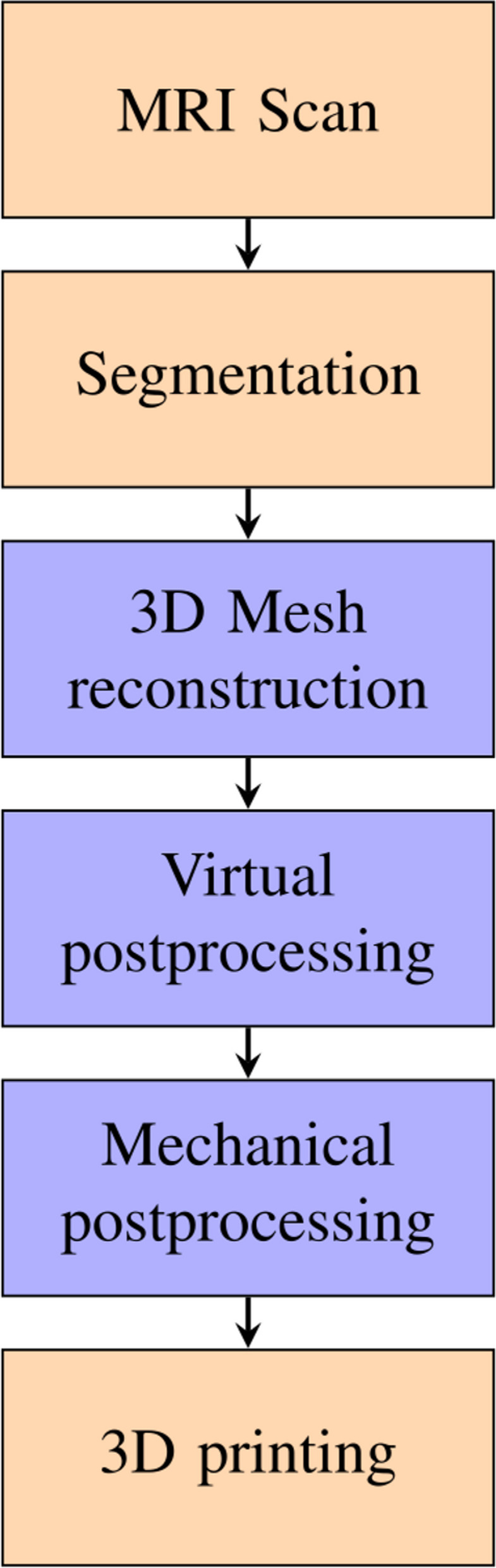
Methods: Magnetic resonance imaging (MRI) of the pelvis was performed on a 3.0 Tesla MRI, including a T2-weighted isotropic 3D sequence. The MRI images were segmented and transferred to virtual 3D models via a custom-built 3D-model generation pipeline and printed by material extrusion. The 3D models were evaluated by all medical specialties involved in patient care of cervical cancer, namely surgeons, radiologists, pathologists and radiation oncologists. Information was obtained from evaluated profession-specific questionnaires which were filled out after inspecting all five models. The questionnaires included multiple-select questions, questions based on Likert scales (1 = "strongly disagree " or "not at all useful " up to 5 = "strongly agree " or "extremely useful ") and dichotomous questions ("Yes" or "No").
Results: Surgeons rated the models as useful during surgery (4.0 out of 5) and for patient communication (4.7 out of 5). Furthermore, they believed that the models had the potential to revise the patients' treatment plan (3.7 out of 5). Pathologists evaluated with mean ratings of 3.0 out of 5 for the usefulness of the models in diagnostic reporting and macroscopic evaluation. Radiologist acknowledged the possibility of providing additional information compared to imaging alone (3.7 out of 5). Radiation oncologists strongly supported the concept by rating the models highly for understanding patient-specific pathological characteristics (4.3 out of 5), assisting interprofessional communication (mean 4.3 out of 5) and communication with patients (4.7 out of 5). They also found the models useful for improving radiotherapy treatment planning (4.3 out of 5).
Conclusion: The study revealed that the 3D printed models were generally well-received by all medical disciplines, with radiation oncologists showing particularly strong support. Addressing the concerns and tailoring the use of 3D models to the specific needs of each medical speciality will be essential for realizing their full potential in clinical practice.


 分享
分享

 求助内容:
求助内容: 应助结果提醒方式:
 扫码关注我们
