Lin Yang, Haiwei Zhang, Jiexin Sheng, Meng Wang, Yaliang Liu, Min Xu, Xiao Yang, Bo Wang, Xiaolong He, Lei Gao, Chao Zheng
{"title":"对比增强增强技术提高了从 80 kVp 脑 CT 灌注数据中提取的 CT 血管造影的图像质量。","authors":"Lin Yang, Haiwei Zhang, Jiexin Sheng, Meng Wang, Yaliang Liu, Min Xu, Xiao Yang, Bo Wang, Xiaolong He, Lei Gao, Chao Zheng","doi":"10.1186/s12880-024-01373-7","DOIUrl":null,"url":null,"abstract":"<p><strong>Rationale and objective: </strong>To investigate the impact of the contrast enhancement boost (CE-boost) technique on the image quality of CT angiography (CTA) derived from 80-kVp cerebral CT perfusion (CTP) data, and to compare it with conventional CTA<sub>peak</sub> as well as other currently employed methods for enhancing CTA images, such as CTA<sub>tMIP</sub> and CTA<sub>tAve</sub> extracted from CTP.</p><p><strong>Materials and methods: </strong>The data of forty-seven patients who underwent CTP at 80 kVp were retrospectively collected. Four sets of images: CTA<sub>peak</sub>, CTA<sub>tMIP</sub>, CTA<sub>tAve</sub>, and CE-boost images. The CTA<sub>peak</sub> image represents the arterial phase at its peak value, captured as a single time point. CTA<sub>tMIP</sub> and CTA<sub>tAve</sub> are 4D CTA images that provide maximum density projection and average images from the three most prominent time points. CE-boost is a postprocessing technique used to enhance contrast in the arterial phase at its peak value. We compared the average CT value, standard deviation (SD), signal-to-noise ratio (SNR), and contrast-to-noise ratio (CNR) of the internal carotid artery (ICA) and basilar artery (BA) among the four groups. Image quality was evaluated using a 5-point scale.</p><p><strong>Results: </strong>The CE-boost demonstrated and CNR in the ICA and BA (all p < 0.001). Compared with the other three CTA reconstructed images, the CE-boost images had the best subjective image quality, with the highest scores of 4.77 ± 0.43 and 4.87 ± 0.34 for each reader (all p < 0.001).</p><p><strong>Conclusion: </strong>Compared with other currently used techniques,CE-boost enhances the image quality of CTA derived from 80-kVp CTP data, leading to improved visualization of intracranial arteries.</p>","PeriodicalId":9020,"journal":{"name":"BMC Medical Imaging","volume":"24 1","pages":"193"},"PeriodicalIF":3.9000,"publicationDate":"2024-07-30","publicationTypes":"Journal Article","fieldsOfStudy":null,"isOpenAccess":false,"openAccessPdf":"https://www.ncbi.nlm.nih.gov/pmc/articles/PMC11290218/pdf/","citationCount":"0","resultStr":"{\"title\":\"Contrast enhancement boost improves the image quality of CT angiography derived from 80-kVp cerebral CT perfusion data.\",\"authors\":\"Lin Yang, Haiwei Zhang, Jiexin Sheng, Meng Wang, Yaliang Liu, Min Xu, Xiao Yang, Bo Wang, Xiaolong He, Lei Gao, Chao Zheng\",\"doi\":\"10.1186/s12880-024-01373-7\",\"DOIUrl\":null,\"url\":null,\"abstract\":\"<p><strong>Rationale and objective: </strong>To investigate the impact of the contrast enhancement boost (CE-boost) technique on the image quality of CT angiography (CTA) derived from 80-kVp cerebral CT perfusion (CTP) data, and to compare it with conventional CTA<sub>peak</sub> as well as other currently employed methods for enhancing CTA images, such as CTA<sub>tMIP</sub> and CTA<sub>tAve</sub> extracted from CTP.</p><p><strong>Materials and methods: </strong>The data of forty-seven patients who underwent CTP at 80 kVp were retrospectively collected. Four sets of images: CTA<sub>peak</sub>, CTA<sub>tMIP</sub>, CTA<sub>tAve</sub>, and CE-boost images. The CTA<sub>peak</sub> image represents the arterial phase at its peak value, captured as a single time point. CTA<sub>tMIP</sub> and CTA<sub>tAve</sub> are 4D CTA images that provide maximum density projection and average images from the three most prominent time points. CE-boost is a postprocessing technique used to enhance contrast in the arterial phase at its peak value. We compared the average CT value, standard deviation (SD), signal-to-noise ratio (SNR), and contrast-to-noise ratio (CNR) of the internal carotid artery (ICA) and basilar artery (BA) among the four groups. Image quality was evaluated using a 5-point scale.</p><p><strong>Results: </strong>The CE-boost demonstrated and CNR in the ICA and BA (all p < 0.001). Compared with the other three CTA reconstructed images, the CE-boost images had the best subjective image quality, with the highest scores of 4.77 ± 0.43 and 4.87 ± 0.34 for each reader (all p < 0.001).</p><p><strong>Conclusion: </strong>Compared with other currently used techniques,CE-boost enhances the image quality of CTA derived from 80-kVp CTP data, leading to improved visualization of intracranial arteries.</p>\",\"PeriodicalId\":9020,\"journal\":{\"name\":\"BMC Medical Imaging\",\"volume\":\"24 1\",\"pages\":\"193\"},\"PeriodicalIF\":3.9000,\"publicationDate\":\"2024-07-30\",\"publicationTypes\":\"Journal Article\",\"fieldsOfStudy\":null,\"isOpenAccess\":false,\"openAccessPdf\":\"https://www.ncbi.nlm.nih.gov/pmc/articles/PMC11290218/pdf/\",\"citationCount\":\"0\",\"resultStr\":null,\"platform\":\"Semanticscholar\",\"paperid\":null,\"PeriodicalName\":\"BMC Medical Imaging\",\"FirstCategoryId\":\"3\",\"ListUrlMain\":\"https://doi.org/10.1186/s12880-024-01373-7\",\"RegionNum\":3,\"RegionCategory\":\"医学\",\"ArticlePicture\":[],\"TitleCN\":null,\"AbstractTextCN\":null,\"PMCID\":null,\"EPubDate\":\"\",\"PubModel\":\"\",\"JCR\":\"Q2\",\"JCRName\":\"RADIOLOGY, NUCLEAR MEDICINE & MEDICAL IMAGING\",\"Score\":null,\"Total\":0}","platform":"Semanticscholar","paperid":null,"PeriodicalName":"BMC Medical Imaging","FirstCategoryId":"3","ListUrlMain":"https://doi.org/10.1186/s12880-024-01373-7","RegionNum":3,"RegionCategory":"医学","ArticlePicture":[],"TitleCN":null,"AbstractTextCN":null,"PMCID":null,"EPubDate":"","PubModel":"","JCR":"Q2","JCRName":"RADIOLOGY, NUCLEAR MEDICINE & MEDICAL IMAGING","Score":null,"Total":0}
Contrast enhancement boost improves the image quality of CT angiography derived from 80-kVp cerebral CT perfusion data.
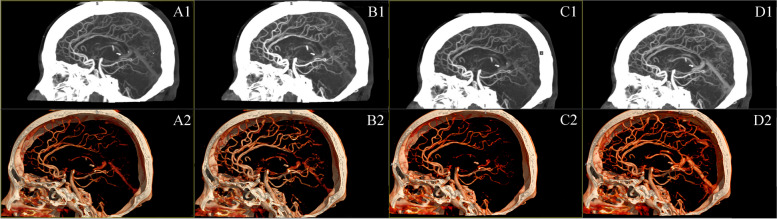
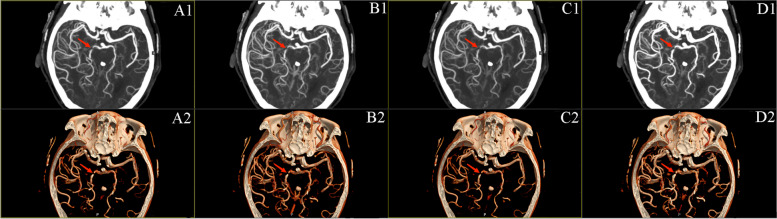
Rationale and objective: To investigate the impact of the contrast enhancement boost (CE-boost) technique on the image quality of CT angiography (CTA) derived from 80-kVp cerebral CT perfusion (CTP) data, and to compare it with conventional CTApeak as well as other currently employed methods for enhancing CTA images, such as CTAtMIP and CTAtAve extracted from CTP.
Materials and methods: The data of forty-seven patients who underwent CTP at 80 kVp were retrospectively collected. Four sets of images: CTApeak, CTAtMIP, CTAtAve, and CE-boost images. The CTApeak image represents the arterial phase at its peak value, captured as a single time point. CTAtMIP and CTAtAve are 4D CTA images that provide maximum density projection and average images from the three most prominent time points. CE-boost is a postprocessing technique used to enhance contrast in the arterial phase at its peak value. We compared the average CT value, standard deviation (SD), signal-to-noise ratio (SNR), and contrast-to-noise ratio (CNR) of the internal carotid artery (ICA) and basilar artery (BA) among the four groups. Image quality was evaluated using a 5-point scale.
Results: The CE-boost demonstrated and CNR in the ICA and BA (all p < 0.001). Compared with the other three CTA reconstructed images, the CE-boost images had the best subjective image quality, with the highest scores of 4.77 ± 0.43 and 4.87 ± 0.34 for each reader (all p < 0.001).
Conclusion: Compared with other currently used techniques,CE-boost enhances the image quality of CTA derived from 80-kVp CTP data, leading to improved visualization of intracranial arteries.
期刊介绍:
BMC Medical Imaging is an open access journal publishing original peer-reviewed research articles in the development, evaluation, and use of imaging techniques and image processing tools to diagnose and manage disease.


 分享
分享
 求助内容:
求助内容: 应助结果提醒方式:
 扫码关注我们
