Hany A Zaki, Bilal Albaroudi, Eman E Shaban, Mohamed Elgassim, Nood Dhafi Almarri, Kaleem Basharat, Ahmed Shaban
{"title":"深静脉血栓 (DVT) 诊断:从急诊护理点超声波 (PoCUS) 技术中汲取灵感:系统综述和荟萃分析。","authors":"Hany A Zaki, Bilal Albaroudi, Eman E Shaban, Mohamed Elgassim, Nood Dhafi Almarri, Kaleem Basharat, Ahmed Shaban","doi":"10.1186/s13089-024-00378-1","DOIUrl":null,"url":null,"abstract":"<p><strong>Background: </strong>The assessment of deep venous thrombosis (DVT) is clinically difficult diagnosis. The \"gold standard test\" for DVT diagnosis is venography; however, various point-of-care ultrasound (POCUS) protocols have been suggested for DVT evaluation in the emergency department.</p><p><strong>Aims: </strong>This review evaluated the role of different POCUS protocols in diagnosing DVT in the emergency department.</p><p><strong>Methods: </strong>A systematic review and meta-analysis was conducted based of PRISMA guideline and registered on PROSEPRO (CRD42023398871). An electronic database search in Embase, PubMed, ScienceDirect, and Google scholar and a manual search were performed to identify eligible studies till February 2023. Quality Assessment of Diagnostic Accuracy Studies tool (QUADAS-2) was used to assess the risk of bias in included studies. Quantitative analysis was carried out using STATA 16 and Review Manager software (RevMan 5.4.1). Sensitivity, specificity of POCUS protocols for DVT diagnosis compared to reference standard test was calculated.</p><p><strong>Results: </strong>Heterogeneity was identified between 26 included studies for review. The pooled sensitivity, specificity, PPV, and NPV for the 2-point POCUS protocol were 92.32% (95% CI: 87.58-97.06), 96.86% (95% CI: 95.09-98.64), 88.41% (95% CI: 82.24-94.58) and 97.25% (95% CI: 95.51-98.99), respectively. Similarly, the pooled sensitivity, specificity, PPV, and NPV for 3-point POCUS were 89.15% (95% CI: 83.24-95.07), 92.71% (95% CI: 89.59-95.83), 81.27% (95% CI: 73.79-88.75), and 95.47% (95% CI: 92.93-98). The data pooled for complete compression ultrasound, and whole-leg duplex ultrasound also resulted in a sensitivity and specificity of 100% (95% CI: 98.21-100) and 97.05% (95% CI: 92.25-100), respectively. On the other hand, the time from triage to DVT diagnosis was significantly shorter for emergency physician-performed POCUS than diagnostic tests performed by radiologists.</p><p><strong>Conclusion: </strong>The diagnostic performance of POCUS protocols performed by emergency physicians was excellent. Combined with the significant reduction in time to diagnosis. POCUS can be used as the first-line imaging tool for DVT diagnosis in the emergency department. We also recommended that attending emergency physicians with POCUS training are present during DVT diagnosis to improve diagnostic performance even though high diagnostic performance is observed even with the minimum training.</p>","PeriodicalId":36911,"journal":{"name":"Ultrasound Journal","volume":"16 1","pages":"37"},"PeriodicalIF":2.9000,"publicationDate":"2024-07-30","publicationTypes":"Journal Article","fieldsOfStudy":null,"isOpenAccess":false,"openAccessPdf":"https://www.ncbi.nlm.nih.gov/pmc/articles/PMC11289207/pdf/","citationCount":"0","resultStr":"{\"title\":\"Deep venous thrombosis (DVT) diagnostics: gleaning insights from point-of-care ultrasound (PoCUS) techniques in emergencies: a systematic review and meta-analysis.\",\"authors\":\"Hany A Zaki, Bilal Albaroudi, Eman E Shaban, Mohamed Elgassim, Nood Dhafi Almarri, Kaleem Basharat, Ahmed Shaban\",\"doi\":\"10.1186/s13089-024-00378-1\",\"DOIUrl\":null,\"url\":null,\"abstract\":\"<p><strong>Background: </strong>The assessment of deep venous thrombosis (DVT) is clinically difficult diagnosis. The \\\"gold standard test\\\" for DVT diagnosis is venography; however, various point-of-care ultrasound (POCUS) protocols have been suggested for DVT evaluation in the emergency department.</p><p><strong>Aims: </strong>This review evaluated the role of different POCUS protocols in diagnosing DVT in the emergency department.</p><p><strong>Methods: </strong>A systematic review and meta-analysis was conducted based of PRISMA guideline and registered on PROSEPRO (CRD42023398871). An electronic database search in Embase, PubMed, ScienceDirect, and Google scholar and a manual search were performed to identify eligible studies till February 2023. Quality Assessment of Diagnostic Accuracy Studies tool (QUADAS-2) was used to assess the risk of bias in included studies. Quantitative analysis was carried out using STATA 16 and Review Manager software (RevMan 5.4.1). Sensitivity, specificity of POCUS protocols for DVT diagnosis compared to reference standard test was calculated.</p><p><strong>Results: </strong>Heterogeneity was identified between 26 included studies for review. The pooled sensitivity, specificity, PPV, and NPV for the 2-point POCUS protocol were 92.32% (95% CI: 87.58-97.06), 96.86% (95% CI: 95.09-98.64), 88.41% (95% CI: 82.24-94.58) and 97.25% (95% CI: 95.51-98.99), respectively. Similarly, the pooled sensitivity, specificity, PPV, and NPV for 3-point POCUS were 89.15% (95% CI: 83.24-95.07), 92.71% (95% CI: 89.59-95.83), 81.27% (95% CI: 73.79-88.75), and 95.47% (95% CI: 92.93-98). The data pooled for complete compression ultrasound, and whole-leg duplex ultrasound also resulted in a sensitivity and specificity of 100% (95% CI: 98.21-100) and 97.05% (95% CI: 92.25-100), respectively. On the other hand, the time from triage to DVT diagnosis was significantly shorter for emergency physician-performed POCUS than diagnostic tests performed by radiologists.</p><p><strong>Conclusion: </strong>The diagnostic performance of POCUS protocols performed by emergency physicians was excellent. Combined with the significant reduction in time to diagnosis. POCUS can be used as the first-line imaging tool for DVT diagnosis in the emergency department. We also recommended that attending emergency physicians with POCUS training are present during DVT diagnosis to improve diagnostic performance even though high diagnostic performance is observed even with the minimum training.</p>\",\"PeriodicalId\":36911,\"journal\":{\"name\":\"Ultrasound Journal\",\"volume\":\"16 1\",\"pages\":\"37\"},\"PeriodicalIF\":2.9000,\"publicationDate\":\"2024-07-30\",\"publicationTypes\":\"Journal Article\",\"fieldsOfStudy\":null,\"isOpenAccess\":false,\"openAccessPdf\":\"https://www.ncbi.nlm.nih.gov/pmc/articles/PMC11289207/pdf/\",\"citationCount\":\"0\",\"resultStr\":null,\"platform\":\"Semanticscholar\",\"paperid\":null,\"PeriodicalName\":\"Ultrasound Journal\",\"FirstCategoryId\":\"1085\",\"ListUrlMain\":\"https://doi.org/10.1186/s13089-024-00378-1\",\"RegionNum\":0,\"RegionCategory\":null,\"ArticlePicture\":[],\"TitleCN\":null,\"AbstractTextCN\":null,\"PMCID\":null,\"EPubDate\":\"\",\"PubModel\":\"\",\"JCR\":\"Q2\",\"JCRName\":\"Medicine\",\"Score\":null,\"Total\":0}","platform":"Semanticscholar","paperid":null,"PeriodicalName":"Ultrasound Journal","FirstCategoryId":"1085","ListUrlMain":"https://doi.org/10.1186/s13089-024-00378-1","RegionNum":0,"RegionCategory":null,"ArticlePicture":[],"TitleCN":null,"AbstractTextCN":null,"PMCID":null,"EPubDate":"","PubModel":"","JCR":"Q2","JCRName":"Medicine","Score":null,"Total":0}
Deep venous thrombosis (DVT) diagnostics: gleaning insights from point-of-care ultrasound (PoCUS) techniques in emergencies: a systematic review and meta-analysis.
Background: The assessment of deep venous thrombosis (DVT) is clinically difficult diagnosis. The "gold standard test" for DVT diagnosis is venography; however, various point-of-care ultrasound (POCUS) protocols have been suggested for DVT evaluation in the emergency department.
Aims: This review evaluated the role of different POCUS protocols in diagnosing DVT in the emergency department.
Methods: A systematic review and meta-analysis was conducted based of PRISMA guideline and registered on PROSEPRO (CRD42023398871). An electronic database search in Embase, PubMed, ScienceDirect, and Google scholar and a manual search were performed to identify eligible studies till February 2023. Quality Assessment of Diagnostic Accuracy Studies tool (QUADAS-2) was used to assess the risk of bias in included studies. Quantitative analysis was carried out using STATA 16 and Review Manager software (RevMan 5.4.1). Sensitivity, specificity of POCUS protocols for DVT diagnosis compared to reference standard test was calculated.
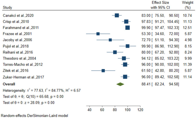
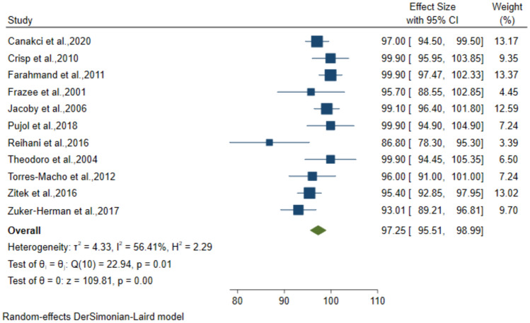
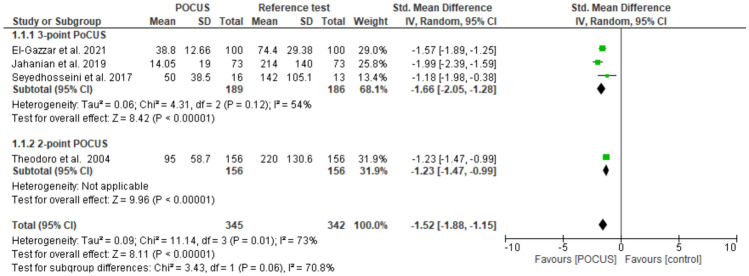
Results: Heterogeneity was identified between 26 included studies for review. The pooled sensitivity, specificity, PPV, and NPV for the 2-point POCUS protocol were 92.32% (95% CI: 87.58-97.06), 96.86% (95% CI: 95.09-98.64), 88.41% (95% CI: 82.24-94.58) and 97.25% (95% CI: 95.51-98.99), respectively. Similarly, the pooled sensitivity, specificity, PPV, and NPV for 3-point POCUS were 89.15% (95% CI: 83.24-95.07), 92.71% (95% CI: 89.59-95.83), 81.27% (95% CI: 73.79-88.75), and 95.47% (95% CI: 92.93-98). The data pooled for complete compression ultrasound, and whole-leg duplex ultrasound also resulted in a sensitivity and specificity of 100% (95% CI: 98.21-100) and 97.05% (95% CI: 92.25-100), respectively. On the other hand, the time from triage to DVT diagnosis was significantly shorter for emergency physician-performed POCUS than diagnostic tests performed by radiologists.
Conclusion: The diagnostic performance of POCUS protocols performed by emergency physicians was excellent. Combined with the significant reduction in time to diagnosis. POCUS can be used as the first-line imaging tool for DVT diagnosis in the emergency department. We also recommended that attending emergency physicians with POCUS training are present during DVT diagnosis to improve diagnostic performance even though high diagnostic performance is observed even with the minimum training.


 分享
分享
 求助内容:
求助内容: 应助结果提醒方式:
 扫码关注我们
