Manmeet S. Dhiman, Taylor J. Bader, Dragana Ponjevic, Paul T. Salo, David A. Hart, Ganesh Swamy, John R. Matyas, Neil A. Duncan
{"title":"用胶原杂交肽量化椎间盘退行性病变患者纤维环的胶原完整性。","authors":"Manmeet S. Dhiman, Taylor J. Bader, Dragana Ponjevic, Paul T. Salo, David A. Hart, Ganesh Swamy, John R. Matyas, Neil A. Duncan","doi":"10.1002/jsp2.1359","DOIUrl":null,"url":null,"abstract":"<div>\n \n \n <section>\n \n <h3> Introduction</h3>\n \n <p>Degenerative disc disease (DDD) is accompanied by structural changes in the intervertebral discs (IVD). Extra-cellular matrix degradation of the annulus fibrosus (AF) has been linked with degeneration of the IVD. Collagen is a vital component of the IVD. Collagen hybridizing peptide (CHP) is an engineered protein that binds to degraded collagen, which we used to quantify collagen damage in AF. This method was used to compare AF samples obtained from donors with no DDD to AF samples from patients undergoing surgery for symptomatic DDD.</p>\n </section>\n \n <section>\n \n <h3> Methods</h3>\n \n <p>Fresh AF tissue was embedded in an optimal cutting temperature compound and cryosectioned at a thickness of 8 μm. Hematoxylin and Eosin staining was performed on sections for general histomorphological assessment. Serial sections were stained with Cy3-conjugated CHP and the mean fluorescence intensity and areal fraction of Cy3-positive staining were averaged for three regions of interest (ROI) on each CHP-stained section.</p>\n </section>\n \n <section>\n \n <h3> Results</h3>\n \n <p>Increases in mean fluorescence intensity (<i>p</i> = 0.0004) and percentage of positively stained area (<i>p</i> = 0.00008) with CHP were detected in DDD samples compared to the non-DDD samples. Significant correlations were observed between mean fluorescence intensity and percentage of positively stained area for both non-DDD (<i>R</i> = 0.98, <i>p</i> = 5E-8) and DDD (<i>R</i> = 0.79, <i>p</i> = 0.0012) samples. No significant differences were detected between sex and the lumbar disc level subgroups of the non-DDD and DDD groups. Only tissue pathology (non-DDD versus DDD) influenced the measured parameters. No three-way interactions between tissue pathology, sex, and lumbar disc level were observed.</p>\n </section>\n \n <section>\n \n <h3> Discussion and Conclusions</h3>\n \n <p>These findings suggest that AF collagen degradation is greater in DDD samples compared to non-DDD samples, as evidenced by the increased CHP staining. Strong positive correlations between the two measured parameters suggest that when collagen degradation occurs, it is detected by this technique and is widespread throughout the tissue. This study provides new insights into the structural alterations associated with collagen degradation in the AF that occur during DDD.</p>\n </section>\n </div>","PeriodicalId":14876,"journal":{"name":"JOR Spine","volume":"7 3","pages":""},"PeriodicalIF":3.9000,"publicationDate":"2024-07-31","publicationTypes":"Journal Article","fieldsOfStudy":null,"isOpenAccess":false,"openAccessPdf":"https://www.ncbi.nlm.nih.gov/pmc/articles/PMC11291301/pdf/","citationCount":"0","resultStr":"{\"title\":\"Collagen integrity of the annulus fibrosus in degenerative disc disease individuals quantified with collagen hybridizing peptide\",\"authors\":\"Manmeet S. Dhiman, Taylor J. Bader, Dragana Ponjevic, Paul T. Salo, David A. Hart, Ganesh Swamy, John R. Matyas, Neil A. Duncan\",\"doi\":\"10.1002/jsp2.1359\",\"DOIUrl\":null,\"url\":null,\"abstract\":\"<div>\\n \\n \\n <section>\\n \\n <h3> Introduction</h3>\\n \\n <p>Degenerative disc disease (DDD) is accompanied by structural changes in the intervertebral discs (IVD). Extra-cellular matrix degradation of the annulus fibrosus (AF) has been linked with degeneration of the IVD. Collagen is a vital component of the IVD. Collagen hybridizing peptide (CHP) is an engineered protein that binds to degraded collagen, which we used to quantify collagen damage in AF. This method was used to compare AF samples obtained from donors with no DDD to AF samples from patients undergoing surgery for symptomatic DDD.</p>\\n </section>\\n \\n <section>\\n \\n <h3> Methods</h3>\\n \\n <p>Fresh AF tissue was embedded in an optimal cutting temperature compound and cryosectioned at a thickness of 8 μm. Hematoxylin and Eosin staining was performed on sections for general histomorphological assessment. Serial sections were stained with Cy3-conjugated CHP and the mean fluorescence intensity and areal fraction of Cy3-positive staining were averaged for three regions of interest (ROI) on each CHP-stained section.</p>\\n </section>\\n \\n <section>\\n \\n <h3> Results</h3>\\n \\n <p>Increases in mean fluorescence intensity (<i>p</i> = 0.0004) and percentage of positively stained area (<i>p</i> = 0.00008) with CHP were detected in DDD samples compared to the non-DDD samples. Significant correlations were observed between mean fluorescence intensity and percentage of positively stained area for both non-DDD (<i>R</i> = 0.98, <i>p</i> = 5E-8) and DDD (<i>R</i> = 0.79, <i>p</i> = 0.0012) samples. No significant differences were detected between sex and the lumbar disc level subgroups of the non-DDD and DDD groups. Only tissue pathology (non-DDD versus DDD) influenced the measured parameters. No three-way interactions between tissue pathology, sex, and lumbar disc level were observed.</p>\\n </section>\\n \\n <section>\\n \\n <h3> Discussion and Conclusions</h3>\\n \\n <p>These findings suggest that AF collagen degradation is greater in DDD samples compared to non-DDD samples, as evidenced by the increased CHP staining. Strong positive correlations between the two measured parameters suggest that when collagen degradation occurs, it is detected by this technique and is widespread throughout the tissue. This study provides new insights into the structural alterations associated with collagen degradation in the AF that occur during DDD.</p>\\n </section>\\n </div>\",\"PeriodicalId\":14876,\"journal\":{\"name\":\"JOR Spine\",\"volume\":\"7 3\",\"pages\":\"\"},\"PeriodicalIF\":3.9000,\"publicationDate\":\"2024-07-31\",\"publicationTypes\":\"Journal Article\",\"fieldsOfStudy\":null,\"isOpenAccess\":false,\"openAccessPdf\":\"https://www.ncbi.nlm.nih.gov/pmc/articles/PMC11291301/pdf/\",\"citationCount\":\"0\",\"resultStr\":null,\"platform\":\"Semanticscholar\",\"paperid\":null,\"PeriodicalName\":\"JOR Spine\",\"FirstCategoryId\":\"3\",\"ListUrlMain\":\"https://onlinelibrary.wiley.com/doi/10.1002/jsp2.1359\",\"RegionNum\":3,\"RegionCategory\":\"医学\",\"ArticlePicture\":[],\"TitleCN\":null,\"AbstractTextCN\":null,\"PMCID\":null,\"EPubDate\":\"\",\"PubModel\":\"\",\"JCR\":\"Q1\",\"JCRName\":\"ORTHOPEDICS\",\"Score\":null,\"Total\":0}","platform":"Semanticscholar","paperid":null,"PeriodicalName":"JOR Spine","FirstCategoryId":"3","ListUrlMain":"https://onlinelibrary.wiley.com/doi/10.1002/jsp2.1359","RegionNum":3,"RegionCategory":"医学","ArticlePicture":[],"TitleCN":null,"AbstractTextCN":null,"PMCID":null,"EPubDate":"","PubModel":"","JCR":"Q1","JCRName":"ORTHOPEDICS","Score":null,"Total":0}
Collagen integrity of the annulus fibrosus in degenerative disc disease individuals quantified with collagen hybridizing peptide
Introduction
Degenerative disc disease (DDD) is accompanied by structural changes in the intervertebral discs (IVD). Extra-cellular matrix degradation of the annulus fibrosus (AF) has been linked with degeneration of the IVD. Collagen is a vital component of the IVD. Collagen hybridizing peptide (CHP) is an engineered protein that binds to degraded collagen, which we used to quantify collagen damage in AF. This method was used to compare AF samples obtained from donors with no DDD to AF samples from patients undergoing surgery for symptomatic DDD.
Methods
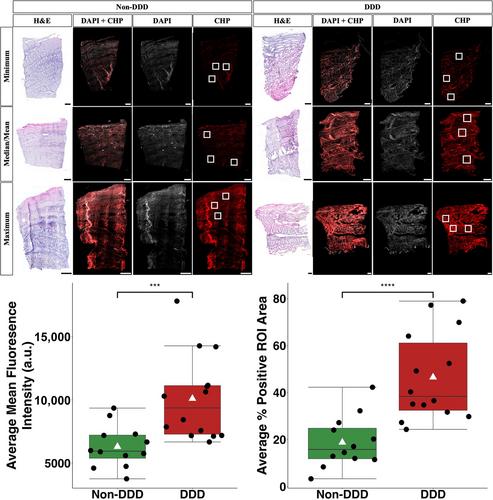
Fresh AF tissue was embedded in an optimal cutting temperature compound and cryosectioned at a thickness of 8 μm. Hematoxylin and Eosin staining was performed on sections for general histomorphological assessment. Serial sections were stained with Cy3-conjugated CHP and the mean fluorescence intensity and areal fraction of Cy3-positive staining were averaged for three regions of interest (ROI) on each CHP-stained section.
Results
Increases in mean fluorescence intensity (p = 0.0004) and percentage of positively stained area (p = 0.00008) with CHP were detected in DDD samples compared to the non-DDD samples. Significant correlations were observed between mean fluorescence intensity and percentage of positively stained area for both non-DDD (R = 0.98, p = 5E-8) and DDD (R = 0.79, p = 0.0012) samples. No significant differences were detected between sex and the lumbar disc level subgroups of the non-DDD and DDD groups. Only tissue pathology (non-DDD versus DDD) influenced the measured parameters. No three-way interactions between tissue pathology, sex, and lumbar disc level were observed.
Discussion and Conclusions
These findings suggest that AF collagen degradation is greater in DDD samples compared to non-DDD samples, as evidenced by the increased CHP staining. Strong positive correlations between the two measured parameters suggest that when collagen degradation occurs, it is detected by this technique and is widespread throughout the tissue. This study provides new insights into the structural alterations associated with collagen degradation in the AF that occur during DDD.


 分享
分享
 求助内容:
求助内容: 应助结果提醒方式:
 扫码关注我们
