Yuyang Tang, Sen Yang, Zhen Qiu, Li Guan, Yigui Wang, Guixin Li, Yuanyu Tu, Lijuan Guo
{"title":"雷帕霉素可减轻H2O2诱导的人皮肤成纤维细胞氧化应激导致的衰老","authors":"Yuyang Tang, Sen Yang, Zhen Qiu, Li Guan, Yigui Wang, Guixin Li, Yuanyu Tu, Lijuan Guo","doi":"10.1007/s13770-024-00660-2","DOIUrl":null,"url":null,"abstract":"<p><strong>Background: </strong>Oxidative stress plays an important role in the skin aging process. Rapamycin has been shown to have anti-aging effects, but its role in oxidative senescence of skin cells remains unclear. The aim of this study was to explore the effect of rapamycin on oxidative stress-induced skin cell senescence and to illustrate the mechanism.</p><p><strong>Methods: </strong>Primary human skin fibroblasts (HSFs) were extracted and a model of H<sub>2</sub>O<sub>2</sub>-induced oxidative senescence was constructed, and the effects of rapamycin on their value-added and migratory capacities were detected by CCK-8 and scratch assays. SA-β-gal was utilized to detect senescence, oxidatively closely related factors were also assessed. Gene and protein expressions of senescence, oxidative, and autophagy were detected by western blotting and quantitative-PCR. The data were analyzed by one-way analysis of variance.</p><p><strong>Results: </strong>Rapamycin (0.1 nmol/L for 48 h) promoted the proliferative and migration of H<sub>2</sub>O<sub>2</sub>-treated HSFs (p < 0.05), decreased senescent phenotypes SA-β-gal staining and the expression of P53, and MMP-1 proteins, and increased the expression level of COL1A-1 (p < 0.001). Rapamycin also enhanced the activities of SOD and HO-1, and effectively removed intracellular ROS, MDA levels (p < 0.05), in addition, autophagy-related proteins and genes were significantly elevated after rapamycin pretreatment (p < 0.001). Rapamycin upregulated the autophagy pathway to exert its protective effects.</p><p><strong>Conclusion: </strong>Our findings indicate that rapamycin shields HSFs from H<sub>2</sub>O<sub>2</sub>-induced oxidative damage, the mechanism is related to the reduction of intracellular peroxidation and upregulation of autophagy pathway. Therefore, rapamycin has the potential to be useful in the investigation and prevention of signs of aging and oxidative stress.</p>","PeriodicalId":23126,"journal":{"name":"Tissue engineering and regenerative medicine","volume":" ","pages":"1049-1059"},"PeriodicalIF":5.1000,"publicationDate":"2024-10-01","publicationTypes":"Journal Article","fieldsOfStudy":null,"isOpenAccess":false,"openAccessPdf":"https://www.ncbi.nlm.nih.gov/pmc/articles/PMC11416443/pdf/","citationCount":"0","resultStr":"{\"title\":\"Rapamycin Attenuates H<sub>2</sub>O<sub>2</sub>-Induced Oxidative Stress-Related Senescence in Human Skin Fibroblasts.\",\"authors\":\"Yuyang Tang, Sen Yang, Zhen Qiu, Li Guan, Yigui Wang, Guixin Li, Yuanyu Tu, Lijuan Guo\",\"doi\":\"10.1007/s13770-024-00660-2\",\"DOIUrl\":null,\"url\":null,\"abstract\":\"<p><strong>Background: </strong>Oxidative stress plays an important role in the skin aging process. Rapamycin has been shown to have anti-aging effects, but its role in oxidative senescence of skin cells remains unclear. The aim of this study was to explore the effect of rapamycin on oxidative stress-induced skin cell senescence and to illustrate the mechanism.</p><p><strong>Methods: </strong>Primary human skin fibroblasts (HSFs) were extracted and a model of H<sub>2</sub>O<sub>2</sub>-induced oxidative senescence was constructed, and the effects of rapamycin on their value-added and migratory capacities were detected by CCK-8 and scratch assays. SA-β-gal was utilized to detect senescence, oxidatively closely related factors were also assessed. Gene and protein expressions of senescence, oxidative, and autophagy were detected by western blotting and quantitative-PCR. The data were analyzed by one-way analysis of variance.</p><p><strong>Results: </strong>Rapamycin (0.1 nmol/L for 48 h) promoted the proliferative and migration of H<sub>2</sub>O<sub>2</sub>-treated HSFs (p < 0.05), decreased senescent phenotypes SA-β-gal staining and the expression of P53, and MMP-1 proteins, and increased the expression level of COL1A-1 (p < 0.001). Rapamycin also enhanced the activities of SOD and HO-1, and effectively removed intracellular ROS, MDA levels (p < 0.05), in addition, autophagy-related proteins and genes were significantly elevated after rapamycin pretreatment (p < 0.001). Rapamycin upregulated the autophagy pathway to exert its protective effects.</p><p><strong>Conclusion: </strong>Our findings indicate that rapamycin shields HSFs from H<sub>2</sub>O<sub>2</sub>-induced oxidative damage, the mechanism is related to the reduction of intracellular peroxidation and upregulation of autophagy pathway. Therefore, rapamycin has the potential to be useful in the investigation and prevention of signs of aging and oxidative stress.</p>\",\"PeriodicalId\":23126,\"journal\":{\"name\":\"Tissue engineering and regenerative medicine\",\"volume\":\" \",\"pages\":\"1049-1059\"},\"PeriodicalIF\":5.1000,\"publicationDate\":\"2024-10-01\",\"publicationTypes\":\"Journal Article\",\"fieldsOfStudy\":null,\"isOpenAccess\":false,\"openAccessPdf\":\"https://www.ncbi.nlm.nih.gov/pmc/articles/PMC11416443/pdf/\",\"citationCount\":\"0\",\"resultStr\":null,\"platform\":\"Semanticscholar\",\"paperid\":null,\"PeriodicalName\":\"Tissue engineering and regenerative medicine\",\"FirstCategoryId\":\"5\",\"ListUrlMain\":\"https://doi.org/10.1007/s13770-024-00660-2\",\"RegionNum\":4,\"RegionCategory\":\"医学\",\"ArticlePicture\":[],\"TitleCN\":null,\"AbstractTextCN\":null,\"PMCID\":null,\"EPubDate\":\"2024/8/2 0:00:00\",\"PubModel\":\"Epub\",\"JCR\":\"Q2\",\"JCRName\":\"CELL & TISSUE ENGINEERING\",\"Score\":null,\"Total\":0}","platform":"Semanticscholar","paperid":null,"PeriodicalName":"Tissue engineering and regenerative medicine","FirstCategoryId":"5","ListUrlMain":"https://doi.org/10.1007/s13770-024-00660-2","RegionNum":4,"RegionCategory":"医学","ArticlePicture":[],"TitleCN":null,"AbstractTextCN":null,"PMCID":null,"EPubDate":"2024/8/2 0:00:00","PubModel":"Epub","JCR":"Q2","JCRName":"CELL & TISSUE ENGINEERING","Score":null,"Total":0}
Rapamycin Attenuates H2O2-Induced Oxidative Stress-Related Senescence in Human Skin Fibroblasts.
Background: Oxidative stress plays an important role in the skin aging process. Rapamycin has been shown to have anti-aging effects, but its role in oxidative senescence of skin cells remains unclear. The aim of this study was to explore the effect of rapamycin on oxidative stress-induced skin cell senescence and to illustrate the mechanism.
Methods: Primary human skin fibroblasts (HSFs) were extracted and a model of H2O2-induced oxidative senescence was constructed, and the effects of rapamycin on their value-added and migratory capacities were detected by CCK-8 and scratch assays. SA-β-gal was utilized to detect senescence, oxidatively closely related factors were also assessed. Gene and protein expressions of senescence, oxidative, and autophagy were detected by western blotting and quantitative-PCR. The data were analyzed by one-way analysis of variance.
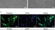
Results: Rapamycin (0.1 nmol/L for 48 h) promoted the proliferative and migration of H2O2-treated HSFs (p < 0.05), decreased senescent phenotypes SA-β-gal staining and the expression of P53, and MMP-1 proteins, and increased the expression level of COL1A-1 (p < 0.001). Rapamycin also enhanced the activities of SOD and HO-1, and effectively removed intracellular ROS, MDA levels (p < 0.05), in addition, autophagy-related proteins and genes were significantly elevated after rapamycin pretreatment (p < 0.001). Rapamycin upregulated the autophagy pathway to exert its protective effects.
Conclusion: Our findings indicate that rapamycin shields HSFs from H2O2-induced oxidative damage, the mechanism is related to the reduction of intracellular peroxidation and upregulation of autophagy pathway. Therefore, rapamycin has the potential to be useful in the investigation and prevention of signs of aging and oxidative stress.
期刊介绍:
Tissue Engineering and Regenerative Medicine (Tissue Eng Regen Med, TERM), the official journal of the Korean Tissue Engineering and Regenerative Medicine Society, is a publication dedicated to providing research- based solutions to issues related to human diseases. This journal publishes articles that report substantial information and original findings on tissue engineering, medical biomaterials, cells therapy, stem cell biology and regenerative medicine.


 分享
分享
 求助内容:
求助内容: 应助结果提醒方式:
 扫码关注我们
