Bing Wu , Tao Zhang , Huabin Chen , Xin Shi , Changbiao Guan , Jianzhong Hu , Hongbin Lu
{"title":"通过低强度脉冲超声刺激预处理的骨髓间充质干细胞提取的外泌体可促进骨-肌腱界面纤维软骨再生,改善肩袖脂肪浸润状况","authors":"Bing Wu , Tao Zhang , Huabin Chen , Xin Shi , Changbiao Guan , Jianzhong Hu , Hongbin Lu","doi":"10.1016/j.jot.2024.07.009","DOIUrl":null,"url":null,"abstract":"<div><h3>Background</h3><p>Fibrovascular scar healing of bone-tendon interface (BTI) instead of functional fibrocartilage regeneration is the main concern associated with unsatisfactory prognosis in rotator cuff repair. Mesenchymal stem cells (MSCs) exosomes have been reported to be a new promising cell-free approach for rotator cuff healing. Whereas, controversies abound in whether exosomes of native MSCs alone can effectively induce chondrogenesis.</p></div><div><h3>Purpose</h3><p>To explore the effect of exosomes derived from low-intensity pulsed ultrasound stimulation (LIPUS)-preconditioned bone marrow mesenchymal stem cells (LIPUS-BMSC-Exos) or un-preconditioned BMSCs (BMSC-Exos) on rotator cuff healing and the underlying mechanism.</p></div><div><h3>Methods</h3><p>C57BL/6 mice underwent unilateral supraspinatus tendon detachment and repair were randomly assigned to saline, BMSCs-Exos or LIPUS-BMSC-Exos injection therapy. Histological, immunofluorescent and biomechanical tests were detected to investigate the effect of exosomes injection on BTI healing and muscle fatty infiltration of the repaired rotator cuff. <em>In vitro</em>, native BMSCs were incubated with BMSC-Exos or LIPUS-BMSC-Exos and then chondrogenic/adipogenic differentiation were observed. Further, quantitative real-time polymerase chain reaction (qRT-PCR) was performed to detect the chondrogenesis/adipogenesis-related miRNA profiles of LIPUS-BMSC-Exos and BMSC-Exos. The chondrogenic/adipogenic potential of the key miRNA was verified through function recover test with its mimic and inhibitor.</p></div><div><h3>Results</h3><p>The results indicated that the biomechanical properties of the supraspinatus tendon-humeral junction were significantly improved in the LIPUS-BMSC-Exos group than that of the BMSCs-Exos group. The LIPUS-BMSC-Exos group also exhibited a higher histological score and more newly regenerated fibrocartilage at the repair site at postoperative 2 and 4 weeks and less fatty infiltration at 4 weeks than the BMSCs-Exos group. <em>In vitro</em>, co-culture of BMSCs with LIPUS-BMSC-Exos could significantly promote BMSCs chondrogenic differentiation and inhibit adipogenic differentiation. Subsequently, qRT-PCR revealed significantly higher enrichment of chondrogenic miRNAs and less enrichment of adipogenic miRNAs in LIPUS-BMSC-Exos compared with BMSC-Exos. Moreover, we demonstrated that this chondrogenesis-inducing potential was primarily attributed to miR-140, one of the most abundant miRNAs in LIPUS-BMSC-Exos.</p></div><div><h3>Conclusion</h3><p>LIPUS-preconditioned BMSC-Exos can effectively promote BTI fibrocartilage regeneration and ameliorate supraspinatus fatty infiltration by positive regulation of pro-chondrogenesis and anti-adipogenesis, which was primarily through delivering miR-140.</p></div><div><h3>The translational potential of this article</h3><p>These findings propose an innovative “LIPUS combined Exosomes strategy” for rotator cuff healing which combines both physiotherapeutic and biotherapeutic advantages. This strategy possesses a good translational potential as a local injection of LIPUS preconditioned BMSC-derived Exos during operation can be not only efficient for promoting fibrocartilage regeneration and ameliorating rotator cuff fatty infiltration, but also time-saving, simple and convenient for patients.</p></div>","PeriodicalId":16636,"journal":{"name":"Journal of Orthopaedic Translation","volume":"48 ","pages":"Pages 89-106"},"PeriodicalIF":5.9000,"publicationDate":"2024-09-01","publicationTypes":"Journal Article","fieldsOfStudy":null,"isOpenAccess":false,"openAccessPdf":"https://www.sciencedirect.com/science/article/pii/S2214031X24000822/pdfft?md5=14328dea7fe6bbbfb76ea8736ec53f5c&pid=1-s2.0-S2214031X24000822-main.pdf","citationCount":"0","resultStr":"{\"title\":\"Exosomes derived from bone marrow mesenchymal stem cell preconditioned by low-intensity pulsed ultrasound stimulation promote bone–tendon interface fibrocartilage regeneration and ameliorate rotator cuff fatty infiltration\",\"authors\":\"Bing Wu , Tao Zhang , Huabin Chen , Xin Shi , Changbiao Guan , Jianzhong Hu , Hongbin Lu\",\"doi\":\"10.1016/j.jot.2024.07.009\",\"DOIUrl\":null,\"url\":null,\"abstract\":\"<div><h3>Background</h3><p>Fibrovascular scar healing of bone-tendon interface (BTI) instead of functional fibrocartilage regeneration is the main concern associated with unsatisfactory prognosis in rotator cuff repair. Mesenchymal stem cells (MSCs) exosomes have been reported to be a new promising cell-free approach for rotator cuff healing. Whereas, controversies abound in whether exosomes of native MSCs alone can effectively induce chondrogenesis.</p></div><div><h3>Purpose</h3><p>To explore the effect of exosomes derived from low-intensity pulsed ultrasound stimulation (LIPUS)-preconditioned bone marrow mesenchymal stem cells (LIPUS-BMSC-Exos) or un-preconditioned BMSCs (BMSC-Exos) on rotator cuff healing and the underlying mechanism.</p></div><div><h3>Methods</h3><p>C57BL/6 mice underwent unilateral supraspinatus tendon detachment and repair were randomly assigned to saline, BMSCs-Exos or LIPUS-BMSC-Exos injection therapy. Histological, immunofluorescent and biomechanical tests were detected to investigate the effect of exosomes injection on BTI healing and muscle fatty infiltration of the repaired rotator cuff. <em>In vitro</em>, native BMSCs were incubated with BMSC-Exos or LIPUS-BMSC-Exos and then chondrogenic/adipogenic differentiation were observed. Further, quantitative real-time polymerase chain reaction (qRT-PCR) was performed to detect the chondrogenesis/adipogenesis-related miRNA profiles of LIPUS-BMSC-Exos and BMSC-Exos. The chondrogenic/adipogenic potential of the key miRNA was verified through function recover test with its mimic and inhibitor.</p></div><div><h3>Results</h3><p>The results indicated that the biomechanical properties of the supraspinatus tendon-humeral junction were significantly improved in the LIPUS-BMSC-Exos group than that of the BMSCs-Exos group. The LIPUS-BMSC-Exos group also exhibited a higher histological score and more newly regenerated fibrocartilage at the repair site at postoperative 2 and 4 weeks and less fatty infiltration at 4 weeks than the BMSCs-Exos group. <em>In vitro</em>, co-culture of BMSCs with LIPUS-BMSC-Exos could significantly promote BMSCs chondrogenic differentiation and inhibit adipogenic differentiation. Subsequently, qRT-PCR revealed significantly higher enrichment of chondrogenic miRNAs and less enrichment of adipogenic miRNAs in LIPUS-BMSC-Exos compared with BMSC-Exos. Moreover, we demonstrated that this chondrogenesis-inducing potential was primarily attributed to miR-140, one of the most abundant miRNAs in LIPUS-BMSC-Exos.</p></div><div><h3>Conclusion</h3><p>LIPUS-preconditioned BMSC-Exos can effectively promote BTI fibrocartilage regeneration and ameliorate supraspinatus fatty infiltration by positive regulation of pro-chondrogenesis and anti-adipogenesis, which was primarily through delivering miR-140.</p></div><div><h3>The translational potential of this article</h3><p>These findings propose an innovative “LIPUS combined Exosomes strategy” for rotator cuff healing which combines both physiotherapeutic and biotherapeutic advantages. This strategy possesses a good translational potential as a local injection of LIPUS preconditioned BMSC-derived Exos during operation can be not only efficient for promoting fibrocartilage regeneration and ameliorating rotator cuff fatty infiltration, but also time-saving, simple and convenient for patients.</p></div>\",\"PeriodicalId\":16636,\"journal\":{\"name\":\"Journal of Orthopaedic Translation\",\"volume\":\"48 \",\"pages\":\"Pages 89-106\"},\"PeriodicalIF\":5.9000,\"publicationDate\":\"2024-09-01\",\"publicationTypes\":\"Journal Article\",\"fieldsOfStudy\":null,\"isOpenAccess\":false,\"openAccessPdf\":\"https://www.sciencedirect.com/science/article/pii/S2214031X24000822/pdfft?md5=14328dea7fe6bbbfb76ea8736ec53f5c&pid=1-s2.0-S2214031X24000822-main.pdf\",\"citationCount\":\"0\",\"resultStr\":null,\"platform\":\"Semanticscholar\",\"paperid\":null,\"PeriodicalName\":\"Journal of Orthopaedic Translation\",\"FirstCategoryId\":\"3\",\"ListUrlMain\":\"https://www.sciencedirect.com/science/article/pii/S2214031X24000822\",\"RegionNum\":1,\"RegionCategory\":\"医学\",\"ArticlePicture\":[],\"TitleCN\":null,\"AbstractTextCN\":null,\"PMCID\":null,\"EPubDate\":\"2024/8/2 0:00:00\",\"PubModel\":\"Epub\",\"JCR\":\"Q1\",\"JCRName\":\"ORTHOPEDICS\",\"Score\":null,\"Total\":0}","platform":"Semanticscholar","paperid":null,"PeriodicalName":"Journal of Orthopaedic Translation","FirstCategoryId":"3","ListUrlMain":"https://www.sciencedirect.com/science/article/pii/S2214031X24000822","RegionNum":1,"RegionCategory":"医学","ArticlePicture":[],"TitleCN":null,"AbstractTextCN":null,"PMCID":null,"EPubDate":"2024/8/2 0:00:00","PubModel":"Epub","JCR":"Q1","JCRName":"ORTHOPEDICS","Score":null,"Total":0}
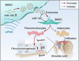
Exosomes derived from bone marrow mesenchymal stem cell preconditioned by low-intensity pulsed ultrasound stimulation promote bone–tendon interface fibrocartilage regeneration and ameliorate rotator cuff fatty infiltration
Background
Fibrovascular scar healing of bone-tendon interface (BTI) instead of functional fibrocartilage regeneration is the main concern associated with unsatisfactory prognosis in rotator cuff repair. Mesenchymal stem cells (MSCs) exosomes have been reported to be a new promising cell-free approach for rotator cuff healing. Whereas, controversies abound in whether exosomes of native MSCs alone can effectively induce chondrogenesis.
Purpose
To explore the effect of exosomes derived from low-intensity pulsed ultrasound stimulation (LIPUS)-preconditioned bone marrow mesenchymal stem cells (LIPUS-BMSC-Exos) or un-preconditioned BMSCs (BMSC-Exos) on rotator cuff healing and the underlying mechanism.
Methods
C57BL/6 mice underwent unilateral supraspinatus tendon detachment and repair were randomly assigned to saline, BMSCs-Exos or LIPUS-BMSC-Exos injection therapy. Histological, immunofluorescent and biomechanical tests were detected to investigate the effect of exosomes injection on BTI healing and muscle fatty infiltration of the repaired rotator cuff. In vitro, native BMSCs were incubated with BMSC-Exos or LIPUS-BMSC-Exos and then chondrogenic/adipogenic differentiation were observed. Further, quantitative real-time polymerase chain reaction (qRT-PCR) was performed to detect the chondrogenesis/adipogenesis-related miRNA profiles of LIPUS-BMSC-Exos and BMSC-Exos. The chondrogenic/adipogenic potential of the key miRNA was verified through function recover test with its mimic and inhibitor.
Results
The results indicated that the biomechanical properties of the supraspinatus tendon-humeral junction were significantly improved in the LIPUS-BMSC-Exos group than that of the BMSCs-Exos group. The LIPUS-BMSC-Exos group also exhibited a higher histological score and more newly regenerated fibrocartilage at the repair site at postoperative 2 and 4 weeks and less fatty infiltration at 4 weeks than the BMSCs-Exos group. In vitro, co-culture of BMSCs with LIPUS-BMSC-Exos could significantly promote BMSCs chondrogenic differentiation and inhibit adipogenic differentiation. Subsequently, qRT-PCR revealed significantly higher enrichment of chondrogenic miRNAs and less enrichment of adipogenic miRNAs in LIPUS-BMSC-Exos compared with BMSC-Exos. Moreover, we demonstrated that this chondrogenesis-inducing potential was primarily attributed to miR-140, one of the most abundant miRNAs in LIPUS-BMSC-Exos.
Conclusion
LIPUS-preconditioned BMSC-Exos can effectively promote BTI fibrocartilage regeneration and ameliorate supraspinatus fatty infiltration by positive regulation of pro-chondrogenesis and anti-adipogenesis, which was primarily through delivering miR-140.
The translational potential of this article
These findings propose an innovative “LIPUS combined Exosomes strategy” for rotator cuff healing which combines both physiotherapeutic and biotherapeutic advantages. This strategy possesses a good translational potential as a local injection of LIPUS preconditioned BMSC-derived Exos during operation can be not only efficient for promoting fibrocartilage regeneration and ameliorating rotator cuff fatty infiltration, but also time-saving, simple and convenient for patients.
期刊介绍:
The Journal of Orthopaedic Translation (JOT) is the official peer-reviewed, open access journal of the Chinese Speaking Orthopaedic Society (CSOS) and the International Chinese Musculoskeletal Research Society (ICMRS). It is published quarterly, in January, April, July and October, by Elsevier.


 分享
分享
 求助内容:
求助内容: 应助结果提醒方式:
 扫码关注我们
