{"title":"根治性前列腺切除术中的术中边缘评估:显微镜是及时冻结还是准备数字解冻?","authors":"Eoin Dinneen, Ricardo Almeida-Magana, Tarek Al-Hammouri, Iona Fernandes, Nikhil Mayor, Larissa Mendes, Mathias Winkler, Anna Silvanto, Aiman Haider, Alex Freeman, Greg Shaw","doi":"10.1111/his.15290","DOIUrl":null,"url":null,"abstract":"<p>Intraoperative frozen section (IFS) is used with the intention to improve functional and oncological outcomes for patients undergoing radical prostatectomy (RP). High resource requirements of IFS techniques such as NeuroSAFE may preclude widespread adoption, even if there are benefits to patients. Recent advances in fresh-tissue microscopic digital imaging technologies may offer an attractive alternative, and there is a growing body of evidence regarding these technologies. In this narrative review, we discuss some of the familiar limitations of IFS and compare these to the attractive counterpoints of modern digital imaging technologies such as the speed and ease of image generation, the locality of equipment within (or near) the operating room, the ability to maintain tissue integrity, and digital transfer of images. Confocal laser microscopy (CLM) is the modality most frequently reported in the literature for margin assessment during RP. We discuss several imitations and obstacles to widespread dissemination of digital imaging technologies. Among these, we consider how the ‘<i>en-face</i>’ margin perspective will challenge urologists and pathologists to understand afresh the meaning of positive margin significance. As a part of this, discussions on how to describe, categorize, react to, and evaluate these technologies are needed to improve patient outcomes. Limitations of this review include its narrative structure and that the evidence base in this field is relatively immature but developing at pace.</p>","PeriodicalId":13219,"journal":{"name":"Histopathology","volume":"85 5","pages":"716-726"},"PeriodicalIF":3.8000,"publicationDate":"2024-08-05","publicationTypes":"Journal Article","fieldsOfStudy":null,"isOpenAccess":false,"openAccessPdf":"https://onlinelibrary.wiley.com/doi/epdf/10.1111/his.15290","citationCount":"0","resultStr":"{\"title\":\"Intraoperative margin assessment during radical prostatectomy: is microscopy frozen in time or ready for digital defrost?\",\"authors\":\"Eoin Dinneen, Ricardo Almeida-Magana, Tarek Al-Hammouri, Iona Fernandes, Nikhil Mayor, Larissa Mendes, Mathias Winkler, Anna Silvanto, Aiman Haider, Alex Freeman, Greg Shaw\",\"doi\":\"10.1111/his.15290\",\"DOIUrl\":null,\"url\":null,\"abstract\":\"<p>Intraoperative frozen section (IFS) is used with the intention to improve functional and oncological outcomes for patients undergoing radical prostatectomy (RP). High resource requirements of IFS techniques such as NeuroSAFE may preclude widespread adoption, even if there are benefits to patients. Recent advances in fresh-tissue microscopic digital imaging technologies may offer an attractive alternative, and there is a growing body of evidence regarding these technologies. In this narrative review, we discuss some of the familiar limitations of IFS and compare these to the attractive counterpoints of modern digital imaging technologies such as the speed and ease of image generation, the locality of equipment within (or near) the operating room, the ability to maintain tissue integrity, and digital transfer of images. Confocal laser microscopy (CLM) is the modality most frequently reported in the literature for margin assessment during RP. We discuss several imitations and obstacles to widespread dissemination of digital imaging technologies. Among these, we consider how the ‘<i>en-face</i>’ margin perspective will challenge urologists and pathologists to understand afresh the meaning of positive margin significance. As a part of this, discussions on how to describe, categorize, react to, and evaluate these technologies are needed to improve patient outcomes. Limitations of this review include its narrative structure and that the evidence base in this field is relatively immature but developing at pace.</p>\",\"PeriodicalId\":13219,\"journal\":{\"name\":\"Histopathology\",\"volume\":\"85 5\",\"pages\":\"716-726\"},\"PeriodicalIF\":3.8000,\"publicationDate\":\"2024-08-05\",\"publicationTypes\":\"Journal Article\",\"fieldsOfStudy\":null,\"isOpenAccess\":false,\"openAccessPdf\":\"https://onlinelibrary.wiley.com/doi/epdf/10.1111/his.15290\",\"citationCount\":\"0\",\"resultStr\":null,\"platform\":\"Semanticscholar\",\"paperid\":null,\"PeriodicalName\":\"Histopathology\",\"FirstCategoryId\":\"3\",\"ListUrlMain\":\"https://onlinelibrary.wiley.com/doi/10.1111/his.15290\",\"RegionNum\":2,\"RegionCategory\":\"医学\",\"ArticlePicture\":[],\"TitleCN\":null,\"AbstractTextCN\":null,\"PMCID\":null,\"EPubDate\":\"\",\"PubModel\":\"\",\"JCR\":\"Q2\",\"JCRName\":\"CELL BIOLOGY\",\"Score\":null,\"Total\":0}","platform":"Semanticscholar","paperid":null,"PeriodicalName":"Histopathology","FirstCategoryId":"3","ListUrlMain":"https://onlinelibrary.wiley.com/doi/10.1111/his.15290","RegionNum":2,"RegionCategory":"医学","ArticlePicture":[],"TitleCN":null,"AbstractTextCN":null,"PMCID":null,"EPubDate":"","PubModel":"","JCR":"Q2","JCRName":"CELL BIOLOGY","Score":null,"Total":0}
引用次数: 0
摘要
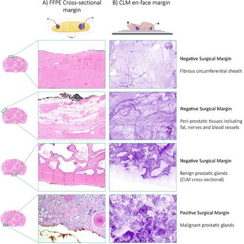
使用术中冰冻切片(IFS)的目的是为了改善根治性前列腺切除术(RP)患者的功能和肿瘤治疗效果。神经前列腺电切术(NeuroSAFE)等术中冷冻切片技术需要大量资源,即使能为患者带来益处,也可能无法得到广泛应用。新鲜组织显微数字成像技术的最新进展可能会提供一种有吸引力的替代方案,而且有关这些技术的证据也在不断增加。在这篇叙述性综述中,我们将讨论人们所熟悉的 IFS 的一些局限性,并将其与现代数字成像技术的诱人之处进行比较,如图像生成的速度和简易性、设备在手术室内(或附近)的位置、保持组织完整性的能力以及图像的数字传输。共焦激光显微镜(CLM)是文献中最常报道的用于 RP 期间边缘评估的方式。我们讨论了数字成像技术在广泛推广过程中的一些模仿和障碍。其中,我们考虑了 "正视 "边缘视角将如何挑战泌尿科医生和病理学家重新理解边缘阳性意义。作为其中的一部分,我们需要讨论如何描述、分类、应对和评估这些技术,以改善患者的治疗效果。本综述的局限性包括其叙述性结构,以及该领域的证据基础相对不成熟,但发展速度较快。
Intraoperative margin assessment during radical prostatectomy: is microscopy frozen in time or ready for digital defrost?
Intraoperative frozen section (IFS) is used with the intention to improve functional and oncological outcomes for patients undergoing radical prostatectomy (RP). High resource requirements of IFS techniques such as NeuroSAFE may preclude widespread adoption, even if there are benefits to patients. Recent advances in fresh-tissue microscopic digital imaging technologies may offer an attractive alternative, and there is a growing body of evidence regarding these technologies. In this narrative review, we discuss some of the familiar limitations of IFS and compare these to the attractive counterpoints of modern digital imaging technologies such as the speed and ease of image generation, the locality of equipment within (or near) the operating room, the ability to maintain tissue integrity, and digital transfer of images. Confocal laser microscopy (CLM) is the modality most frequently reported in the literature for margin assessment during RP. We discuss several imitations and obstacles to widespread dissemination of digital imaging technologies. Among these, we consider how the ‘en-face’ margin perspective will challenge urologists and pathologists to understand afresh the meaning of positive margin significance. As a part of this, discussions on how to describe, categorize, react to, and evaluate these technologies are needed to improve patient outcomes. Limitations of this review include its narrative structure and that the evidence base in this field is relatively immature but developing at pace.
期刊介绍:
Histopathology is an international journal intended to be of practical value to surgical and diagnostic histopathologists, and to investigators of human disease who employ histopathological methods. Our primary purpose is to publish advances in pathology, in particular those applicable to clinical practice and contributing to the better understanding of human disease.


 分享
分享
 求助内容:
求助内容: 应助结果提醒方式:
 扫码关注我们
