Lanette LaComb, Agnidipta Ghosh, Jeffrey B Bonanno, Daniel J Nilson, Alex J Poppel, Lucas Dada, Sean M Cahill, Juan Pablo Maianti, Seiya Kitamura, David Cowburn, Steven C Almo
{"title":"洞察 Mena 的 EVH1 结构域的相互作用格局","authors":"Lanette LaComb, Agnidipta Ghosh, Jeffrey B Bonanno, Daniel J Nilson, Alex J Poppel, Lucas Dada, Sean M Cahill, Juan Pablo Maianti, Seiya Kitamura, David Cowburn, Steven C Almo","doi":"10.1021/acs.biochem.4c00331","DOIUrl":null,"url":null,"abstract":"<p><p>The Enabled/VASP homology 1 (EVH1) domain is a small module that interacts with proline-rich stretches in its ligands and is found in various signaling and scaffolding proteins. Mena, the mammalian homologue of Ena, is involved in diverse actin-associated events, such as membrane dynamics, bacterial motility, and tumor intravasation and extravasation. Two-dimensional (2D) <sup>1</sup>H-<sup>15</sup>N HSQC NMR was used to study Mena EVH1 binding properties, defining the amino acids involved in ligand recognition for the physiological ligands ActA and PCARE, and a synthetic polyproline-inspired small molecule (hereafter inhibitor <b>6c</b>). Chemical shift perturbations indicated that proline-rich segments bind in the conserved EVH1 hydrophobic cleft. The PCARE-derived peptide elicited more perturbations compared to the ActA-derived peptide, consistent with a previous report of a structural alteration in the solvent-exposed β7-β8 loop. Unexpectedly, EVH1 and the proline-rich segment of PTP1B did not exhibit NMR chemical shift perturbations; however, the high-resolution crystal structure implicated the conserved EVH1 hydrophobic cleft in ligand recognition. Intrinsic steady-state fluorescence and fluorescence polarization assays indicate that residues outside the proline-rich segment enhance the ligand affinity for EVH1 (<i>K</i><sub>d</sub> = 3-8 μM). Inhibitor <b>6c</b> displayed tighter binding (<i>K</i><sub>d</sub> ∼ 0.3 μM) and occupies the same EVH1 cleft as physiological ligands. These studies revealed that the EVH1 domain enhances ligand affinity through recognition of residues flanking the proline-rich segments. Additionally, a synthetic inhibitor binds more tightly to the EVH1 domain than natural ligands, occupying the same hydrophobic cleft.</p>","PeriodicalId":28,"journal":{"name":"Biochemistry Biochemistry","volume":" ","pages":"2183-2195"},"PeriodicalIF":3.0000,"publicationDate":"2024-09-03","publicationTypes":"Journal Article","fieldsOfStudy":null,"isOpenAccess":false,"openAccessPdf":"https://www.ncbi.nlm.nih.gov/pmc/articles/PMC12395540/pdf/","citationCount":"0","resultStr":"{\"title\":\"Insights into the Interaction Landscape of the EVH1 Domain of Mena.\",\"authors\":\"Lanette LaComb, Agnidipta Ghosh, Jeffrey B Bonanno, Daniel J Nilson, Alex J Poppel, Lucas Dada, Sean M Cahill, Juan Pablo Maianti, Seiya Kitamura, David Cowburn, Steven C Almo\",\"doi\":\"10.1021/acs.biochem.4c00331\",\"DOIUrl\":null,\"url\":null,\"abstract\":\"<p><p>The Enabled/VASP homology 1 (EVH1) domain is a small module that interacts with proline-rich stretches in its ligands and is found in various signaling and scaffolding proteins. Mena, the mammalian homologue of Ena, is involved in diverse actin-associated events, such as membrane dynamics, bacterial motility, and tumor intravasation and extravasation. Two-dimensional (2D) <sup>1</sup>H-<sup>15</sup>N HSQC NMR was used to study Mena EVH1 binding properties, defining the amino acids involved in ligand recognition for the physiological ligands ActA and PCARE, and a synthetic polyproline-inspired small molecule (hereafter inhibitor <b>6c</b>). Chemical shift perturbations indicated that proline-rich segments bind in the conserved EVH1 hydrophobic cleft. The PCARE-derived peptide elicited more perturbations compared to the ActA-derived peptide, consistent with a previous report of a structural alteration in the solvent-exposed β7-β8 loop. Unexpectedly, EVH1 and the proline-rich segment of PTP1B did not exhibit NMR chemical shift perturbations; however, the high-resolution crystal structure implicated the conserved EVH1 hydrophobic cleft in ligand recognition. Intrinsic steady-state fluorescence and fluorescence polarization assays indicate that residues outside the proline-rich segment enhance the ligand affinity for EVH1 (<i>K</i><sub>d</sub> = 3-8 μM). Inhibitor <b>6c</b> displayed tighter binding (<i>K</i><sub>d</sub> ∼ 0.3 μM) and occupies the same EVH1 cleft as physiological ligands. These studies revealed that the EVH1 domain enhances ligand affinity through recognition of residues flanking the proline-rich segments. Additionally, a synthetic inhibitor binds more tightly to the EVH1 domain than natural ligands, occupying the same hydrophobic cleft.</p>\",\"PeriodicalId\":28,\"journal\":{\"name\":\"Biochemistry Biochemistry\",\"volume\":\" \",\"pages\":\"2183-2195\"},\"PeriodicalIF\":3.0000,\"publicationDate\":\"2024-09-03\",\"publicationTypes\":\"Journal Article\",\"fieldsOfStudy\":null,\"isOpenAccess\":false,\"openAccessPdf\":\"https://www.ncbi.nlm.nih.gov/pmc/articles/PMC12395540/pdf/\",\"citationCount\":\"0\",\"resultStr\":null,\"platform\":\"Semanticscholar\",\"paperid\":null,\"PeriodicalName\":\"Biochemistry Biochemistry\",\"FirstCategoryId\":\"1\",\"ListUrlMain\":\"https://doi.org/10.1021/acs.biochem.4c00331\",\"RegionNum\":3,\"RegionCategory\":\"生物学\",\"ArticlePicture\":[],\"TitleCN\":null,\"AbstractTextCN\":null,\"PMCID\":null,\"EPubDate\":\"2024/8/13 0:00:00\",\"PubModel\":\"Epub\",\"JCR\":\"Q3\",\"JCRName\":\"BIOCHEMISTRY & MOLECULAR BIOLOGY\",\"Score\":null,\"Total\":0}","platform":"Semanticscholar","paperid":null,"PeriodicalName":"Biochemistry Biochemistry","FirstCategoryId":"1","ListUrlMain":"https://doi.org/10.1021/acs.biochem.4c00331","RegionNum":3,"RegionCategory":"生物学","ArticlePicture":[],"TitleCN":null,"AbstractTextCN":null,"PMCID":null,"EPubDate":"2024/8/13 0:00:00","PubModel":"Epub","JCR":"Q3","JCRName":"BIOCHEMISTRY & MOLECULAR BIOLOGY","Score":null,"Total":0}
Insights into the Interaction Landscape of the EVH1 Domain of Mena.
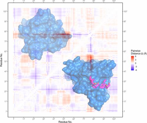
The Enabled/VASP homology 1 (EVH1) domain is a small module that interacts with proline-rich stretches in its ligands and is found in various signaling and scaffolding proteins. Mena, the mammalian homologue of Ena, is involved in diverse actin-associated events, such as membrane dynamics, bacterial motility, and tumor intravasation and extravasation. Two-dimensional (2D) 1H-15N HSQC NMR was used to study Mena EVH1 binding properties, defining the amino acids involved in ligand recognition for the physiological ligands ActA and PCARE, and a synthetic polyproline-inspired small molecule (hereafter inhibitor 6c). Chemical shift perturbations indicated that proline-rich segments bind in the conserved EVH1 hydrophobic cleft. The PCARE-derived peptide elicited more perturbations compared to the ActA-derived peptide, consistent with a previous report of a structural alteration in the solvent-exposed β7-β8 loop. Unexpectedly, EVH1 and the proline-rich segment of PTP1B did not exhibit NMR chemical shift perturbations; however, the high-resolution crystal structure implicated the conserved EVH1 hydrophobic cleft in ligand recognition. Intrinsic steady-state fluorescence and fluorescence polarization assays indicate that residues outside the proline-rich segment enhance the ligand affinity for EVH1 (Kd = 3-8 μM). Inhibitor 6c displayed tighter binding (Kd ∼ 0.3 μM) and occupies the same EVH1 cleft as physiological ligands. These studies revealed that the EVH1 domain enhances ligand affinity through recognition of residues flanking the proline-rich segments. Additionally, a synthetic inhibitor binds more tightly to the EVH1 domain than natural ligands, occupying the same hydrophobic cleft.
期刊介绍:
Biochemistry provides an international forum for publishing exceptional, rigorous, high-impact research across all of biological chemistry. This broad scope includes studies on the chemical, physical, mechanistic, and/or structural basis of biological or cell function, and encompasses the fields of chemical biology, synthetic biology, disease biology, cell biology, nucleic acid biology, neuroscience, structural biology, and biophysics. In addition to traditional Research Articles, Biochemistry also publishes Communications, Viewpoints, and Perspectives, as well as From the Bench articles that report new methods of particular interest to the biological chemistry community.


 分享
分享
 求助内容:
求助内容: 应助结果提醒方式:
 扫码关注我们
