Jae Soo Kim, Mi-Hye Kim, Myeung Ju Kim, Hee Jung Kim
{"title":"甘草查尔酮 A 可减轻 NMDA 诱导的神经毒性。","authors":"Jae Soo Kim, Mi-Hye Kim, Myeung Ju Kim, Hee Jung Kim","doi":"10.1080/19768354.2024.2389823","DOIUrl":null,"url":null,"abstract":"<p><p>This study investigates the effect of Licochalcone A (Lico-A), a flavonoid from licorice roots known for its anti-inflammatory, anti-cancer, and antioxidant properties, on NMDA-induced neurotoxicity in primary cultured rat hippocampal neurons. The study measured cell survival following NMDA and Lico-A exposure, revealing that Lico-A at a 2.5 μg/ml significantly improved cell viability, countering the detrimental effects of NMDA. The study also analyzed synaptic changes by examining both postsynaptic density 95 (PSD95) and synaptophysin-targeted imaging, showing that Lico-A treatment resulted in a significant increase in synaptic puncta, contrasting with the reduction observed under NMDA exposure. Furthermore, levels of phosphorylated mixed lineage kinase domain-like pseudokinase (P-MLKL) and phosphorylated receptor-interacting serine/threonine-protein kinase 3 (P-RIP3), key necroptosis regulators, were measured using Western blotting. The results showed an increase in P-MLKL and P-RIP3 in neurons exposed to NMDA, which was reduced following Lico-A treatment. The response of astrocyte and microglia was also evaluated by immunostaining for glial fibrillary acidic protein (GFAP), ionized calcium-binding adaptor molecule 1 (IBA-1) and tumor necrosis factor alpha (TNF-α). These markers exhibited heightened expression in the NMDA group, which was substantially reduced by Lico-A treatment. These findings suggest that Lico-A has neuroprotective effects against NMDA-induced neurotoxicity, potentially contributing to synaptic preservation, inhibition of neuronal necroptosis, and modulation of glial activation. Therefore, Lico-A shows promise as a neuroprotective agent for conditions associated with NMDA-related neurotoxicity.</p>","PeriodicalId":7804,"journal":{"name":"Animal Cells and Systems","volume":"28 1","pages":"392-400"},"PeriodicalIF":3.6000,"publicationDate":"2024-08-12","publicationTypes":"Journal Article","fieldsOfStudy":null,"isOpenAccess":false,"openAccessPdf":"https://www.ncbi.nlm.nih.gov/pmc/articles/PMC11321100/pdf/","citationCount":"0","resultStr":"{\"title\":\"Licochalcone A attenuates NMDA-induced neurotoxicity.\",\"authors\":\"Jae Soo Kim, Mi-Hye Kim, Myeung Ju Kim, Hee Jung Kim\",\"doi\":\"10.1080/19768354.2024.2389823\",\"DOIUrl\":null,\"url\":null,\"abstract\":\"<p><p>This study investigates the effect of Licochalcone A (Lico-A), a flavonoid from licorice roots known for its anti-inflammatory, anti-cancer, and antioxidant properties, on NMDA-induced neurotoxicity in primary cultured rat hippocampal neurons. The study measured cell survival following NMDA and Lico-A exposure, revealing that Lico-A at a 2.5 μg/ml significantly improved cell viability, countering the detrimental effects of NMDA. The study also analyzed synaptic changes by examining both postsynaptic density 95 (PSD95) and synaptophysin-targeted imaging, showing that Lico-A treatment resulted in a significant increase in synaptic puncta, contrasting with the reduction observed under NMDA exposure. Furthermore, levels of phosphorylated mixed lineage kinase domain-like pseudokinase (P-MLKL) and phosphorylated receptor-interacting serine/threonine-protein kinase 3 (P-RIP3), key necroptosis regulators, were measured using Western blotting. The results showed an increase in P-MLKL and P-RIP3 in neurons exposed to NMDA, which was reduced following Lico-A treatment. The response of astrocyte and microglia was also evaluated by immunostaining for glial fibrillary acidic protein (GFAP), ionized calcium-binding adaptor molecule 1 (IBA-1) and tumor necrosis factor alpha (TNF-α). These markers exhibited heightened expression in the NMDA group, which was substantially reduced by Lico-A treatment. These findings suggest that Lico-A has neuroprotective effects against NMDA-induced neurotoxicity, potentially contributing to synaptic preservation, inhibition of neuronal necroptosis, and modulation of glial activation. Therefore, Lico-A shows promise as a neuroprotective agent for conditions associated with NMDA-related neurotoxicity.</p>\",\"PeriodicalId\":7804,\"journal\":{\"name\":\"Animal Cells and Systems\",\"volume\":\"28 1\",\"pages\":\"392-400\"},\"PeriodicalIF\":3.6000,\"publicationDate\":\"2024-08-12\",\"publicationTypes\":\"Journal Article\",\"fieldsOfStudy\":null,\"isOpenAccess\":false,\"openAccessPdf\":\"https://www.ncbi.nlm.nih.gov/pmc/articles/PMC11321100/pdf/\",\"citationCount\":\"0\",\"resultStr\":null,\"platform\":\"Semanticscholar\",\"paperid\":null,\"PeriodicalName\":\"Animal Cells and Systems\",\"FirstCategoryId\":\"99\",\"ListUrlMain\":\"https://doi.org/10.1080/19768354.2024.2389823\",\"RegionNum\":2,\"RegionCategory\":\"生物学\",\"ArticlePicture\":[],\"TitleCN\":null,\"AbstractTextCN\":null,\"PMCID\":null,\"EPubDate\":\"2024/1/1 0:00:00\",\"PubModel\":\"eCollection\",\"JCR\":\"Q3\",\"JCRName\":\"CELL BIOLOGY\",\"Score\":null,\"Total\":0}","platform":"Semanticscholar","paperid":null,"PeriodicalName":"Animal Cells and Systems","FirstCategoryId":"99","ListUrlMain":"https://doi.org/10.1080/19768354.2024.2389823","RegionNum":2,"RegionCategory":"生物学","ArticlePicture":[],"TitleCN":null,"AbstractTextCN":null,"PMCID":null,"EPubDate":"2024/1/1 0:00:00","PubModel":"eCollection","JCR":"Q3","JCRName":"CELL BIOLOGY","Score":null,"Total":0}
Licochalcone A attenuates NMDA-induced neurotoxicity.
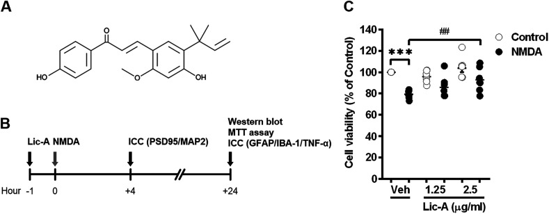
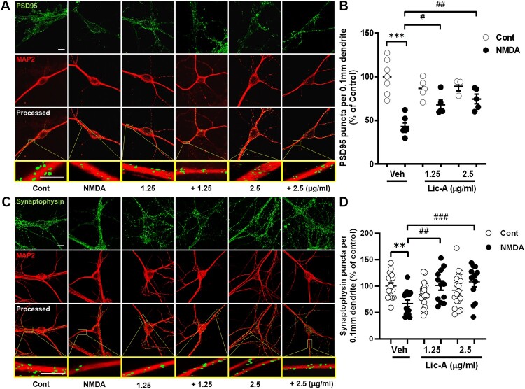
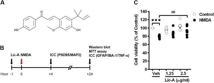
This study investigates the effect of Licochalcone A (Lico-A), a flavonoid from licorice roots known for its anti-inflammatory, anti-cancer, and antioxidant properties, on NMDA-induced neurotoxicity in primary cultured rat hippocampal neurons. The study measured cell survival following NMDA and Lico-A exposure, revealing that Lico-A at a 2.5 μg/ml significantly improved cell viability, countering the detrimental effects of NMDA. The study also analyzed synaptic changes by examining both postsynaptic density 95 (PSD95) and synaptophysin-targeted imaging, showing that Lico-A treatment resulted in a significant increase in synaptic puncta, contrasting with the reduction observed under NMDA exposure. Furthermore, levels of phosphorylated mixed lineage kinase domain-like pseudokinase (P-MLKL) and phosphorylated receptor-interacting serine/threonine-protein kinase 3 (P-RIP3), key necroptosis regulators, were measured using Western blotting. The results showed an increase in P-MLKL and P-RIP3 in neurons exposed to NMDA, which was reduced following Lico-A treatment. The response of astrocyte and microglia was also evaluated by immunostaining for glial fibrillary acidic protein (GFAP), ionized calcium-binding adaptor molecule 1 (IBA-1) and tumor necrosis factor alpha (TNF-α). These markers exhibited heightened expression in the NMDA group, which was substantially reduced by Lico-A treatment. These findings suggest that Lico-A has neuroprotective effects against NMDA-induced neurotoxicity, potentially contributing to synaptic preservation, inhibition of neuronal necroptosis, and modulation of glial activation. Therefore, Lico-A shows promise as a neuroprotective agent for conditions associated with NMDA-related neurotoxicity.
期刊介绍:
Animal Cells and Systems is the official journal of the Korean Society for Integrative Biology. This international, peer-reviewed journal publishes original papers that cover diverse aspects of biological sciences including Bioinformatics and Systems Biology, Developmental Biology, Evolution and Systematic Biology, Population Biology, & Animal Behaviour, Molecular and Cellular Biology, Neurobiology and Immunology, and Translational Medicine.


 分享
分享
 求助内容:
求助内容: 应助结果提醒方式:
 扫码关注我们
