{"title":"交互式多尺度融合:通过跨 IMSM 模型推进脑肿瘤检测。","authors":"Vasanthi Durairaj, Palani Uthirapathy","doi":"10.1007/s10278-024-01222-7","DOIUrl":null,"url":null,"abstract":"<p><p>Multi-modal medical image (MI) fusion assists in generating collaboration images collecting complement features through the distinct images of several conditions. The images help physicians to diagnose disease accurately. Hence, this research proposes a novel multi-modal MI fusion modal named guided filter-based interactive multi-scale and multi-modal transformer (Trans-IMSM) fusion approach to develop high-quality computed tomography-magnetic resonance imaging (CT-MRI) fused images for brain tumor detection. This research utilizes the CT and MRI brain scan dataset to gather the input CT and MRI images. At first, the data preprocessing is carried out to preprocess these input images to improve the image quality and generalization ability for further analysis. Then, these preprocessed CT and MRI are decomposed into detail and base components utilizing the guided filter-based MI decomposition approach. This approach involves two phases: such as acquiring the image guidance and decomposing the images utilizing the guided filter. A canny operator is employed to acquire the image guidance comprising robust edge for CT and MRI images, and the guided filter is applied to decompose the guidance and preprocessed images. Then, by applying the Trans-IMSM model, fuse the detail components, while a weighting approach is used for the base components. The fused detail and base components are subsequently processed through a gated fusion and reconstruction network, and the final fused images for brain tumor detection are generated. Extensive tests are carried out to compute the Trans-IMSM method's efficacy. The evaluation results demonstrated the robustness and effectiveness, achieving an accuracy of 98.64% and an SSIM of 0.94.</p>","PeriodicalId":516858,"journal":{"name":"Journal of imaging informatics in medicine","volume":" ","pages":"757-774"},"PeriodicalIF":0.0000,"publicationDate":"2025-04-01","publicationTypes":"Journal Article","fieldsOfStudy":null,"isOpenAccess":false,"openAccessPdf":"https://www.ncbi.nlm.nih.gov/pmc/articles/PMC11950544/pdf/","citationCount":"0","resultStr":"{\"title\":\"Interactive Multi-scale Fusion: Advancing Brain Tumor Detection Through Trans-IMSM Model.\",\"authors\":\"Vasanthi Durairaj, Palani Uthirapathy\",\"doi\":\"10.1007/s10278-024-01222-7\",\"DOIUrl\":null,\"url\":null,\"abstract\":\"<p><p>Multi-modal medical image (MI) fusion assists in generating collaboration images collecting complement features through the distinct images of several conditions. The images help physicians to diagnose disease accurately. Hence, this research proposes a novel multi-modal MI fusion modal named guided filter-based interactive multi-scale and multi-modal transformer (Trans-IMSM) fusion approach to develop high-quality computed tomography-magnetic resonance imaging (CT-MRI) fused images for brain tumor detection. This research utilizes the CT and MRI brain scan dataset to gather the input CT and MRI images. At first, the data preprocessing is carried out to preprocess these input images to improve the image quality and generalization ability for further analysis. Then, these preprocessed CT and MRI are decomposed into detail and base components utilizing the guided filter-based MI decomposition approach. This approach involves two phases: such as acquiring the image guidance and decomposing the images utilizing the guided filter. A canny operator is employed to acquire the image guidance comprising robust edge for CT and MRI images, and the guided filter is applied to decompose the guidance and preprocessed images. Then, by applying the Trans-IMSM model, fuse the detail components, while a weighting approach is used for the base components. The fused detail and base components are subsequently processed through a gated fusion and reconstruction network, and the final fused images for brain tumor detection are generated. Extensive tests are carried out to compute the Trans-IMSM method's efficacy. The evaluation results demonstrated the robustness and effectiveness, achieving an accuracy of 98.64% and an SSIM of 0.94.</p>\",\"PeriodicalId\":516858,\"journal\":{\"name\":\"Journal of imaging informatics in medicine\",\"volume\":\" \",\"pages\":\"757-774\"},\"PeriodicalIF\":0.0000,\"publicationDate\":\"2025-04-01\",\"publicationTypes\":\"Journal Article\",\"fieldsOfStudy\":null,\"isOpenAccess\":false,\"openAccessPdf\":\"https://www.ncbi.nlm.nih.gov/pmc/articles/PMC11950544/pdf/\",\"citationCount\":\"0\",\"resultStr\":null,\"platform\":\"Semanticscholar\",\"paperid\":null,\"PeriodicalName\":\"Journal of imaging informatics in medicine\",\"FirstCategoryId\":\"1085\",\"ListUrlMain\":\"https://doi.org/10.1007/s10278-024-01222-7\",\"RegionNum\":0,\"RegionCategory\":null,\"ArticlePicture\":[],\"TitleCN\":null,\"AbstractTextCN\":null,\"PMCID\":null,\"EPubDate\":\"2024/8/15 0:00:00\",\"PubModel\":\"Epub\",\"JCR\":\"\",\"JCRName\":\"\",\"Score\":null,\"Total\":0}","platform":"Semanticscholar","paperid":null,"PeriodicalName":"Journal of imaging informatics in medicine","FirstCategoryId":"1085","ListUrlMain":"https://doi.org/10.1007/s10278-024-01222-7","RegionNum":0,"RegionCategory":null,"ArticlePicture":[],"TitleCN":null,"AbstractTextCN":null,"PMCID":null,"EPubDate":"2024/8/15 0:00:00","PubModel":"Epub","JCR":"","JCRName":"","Score":null,"Total":0}
Interactive Multi-scale Fusion: Advancing Brain Tumor Detection Through Trans-IMSM Model.
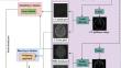
Multi-modal medical image (MI) fusion assists in generating collaboration images collecting complement features through the distinct images of several conditions. The images help physicians to diagnose disease accurately. Hence, this research proposes a novel multi-modal MI fusion modal named guided filter-based interactive multi-scale and multi-modal transformer (Trans-IMSM) fusion approach to develop high-quality computed tomography-magnetic resonance imaging (CT-MRI) fused images for brain tumor detection. This research utilizes the CT and MRI brain scan dataset to gather the input CT and MRI images. At first, the data preprocessing is carried out to preprocess these input images to improve the image quality and generalization ability for further analysis. Then, these preprocessed CT and MRI are decomposed into detail and base components utilizing the guided filter-based MI decomposition approach. This approach involves two phases: such as acquiring the image guidance and decomposing the images utilizing the guided filter. A canny operator is employed to acquire the image guidance comprising robust edge for CT and MRI images, and the guided filter is applied to decompose the guidance and preprocessed images. Then, by applying the Trans-IMSM model, fuse the detail components, while a weighting approach is used for the base components. The fused detail and base components are subsequently processed through a gated fusion and reconstruction network, and the final fused images for brain tumor detection are generated. Extensive tests are carried out to compute the Trans-IMSM method's efficacy. The evaluation results demonstrated the robustness and effectiveness, achieving an accuracy of 98.64% and an SSIM of 0.94.


 分享
分享

 求助内容:
求助内容: 应助结果提醒方式:
 扫码关注我们
