Anne Grethe Jurik, Asta Linauskas, Rosa Marie Kiil
{"title":"通过磁共振成像诊断髂髁炎的特征--系统性文献综述。","authors":"Anne Grethe Jurik, Asta Linauskas, Rosa Marie Kiil","doi":"10.1007/s00256-024-04773-6","DOIUrl":null,"url":null,"abstract":"<p><strong>Objective: </strong>To describe and evaluate the current knowledge of MRI characteristics of osteitis condensans ilii (OCI) in the diagnostics and differentiation of OCI from other conditions.</p><p><strong>Materials and methods: </strong>The databases PubMed, EMBASE, Scopus, and Web of Science were searched from their inception to March 2024 using the search terms \"Magnetic Resonance Imaging\" (MESH term in PubMed) and \"osteitis condensans ilii\" and limited to English language. Two reviewers independently screened titles, abstracts, and full-text eligibility and assessed the risk of bias according to Quality Assessment of Diagnostic Accuracy Studies, QUADAS-2.</p><p><strong>Results: </strong>The search identified 53 records. Case reports, letters/notes, and conference abstracts were excluded, resulting in 24 reports assessed by full-text, 9 research articles, 14 reviews, and a book chapter. Five retrospective research studies were found eligible for the review. Detailed MRI features of OCI were only described in two studies of patients with pain where they encompassed manifest subchondral iliac sclerosis often accompanied by bone marrow edema (BME) located peripheral to the sclerosis and displaying a continuous distribution and frequently accompanied by sacral BME. Erosions were rare and ankylosis did not occur. Fat deposition in the bone marrow was frequent and similar to BME often located to anterior strain-related joint areas. The QUADAS-2 assessments revealed risks of bias in all studies analyzed, especially regarding general applicability of the MRI features.</p><p><strong>Conclusion: </strong>There is a lack of valid data describing characteristic MRI features in general groups of OCI patients with and without pain.</p>","PeriodicalId":21783,"journal":{"name":"Skeletal Radiology","volume":" ","pages":"423-430"},"PeriodicalIF":2.2000,"publicationDate":"2025-03-01","publicationTypes":"Journal Article","fieldsOfStudy":null,"isOpenAccess":false,"openAccessPdf":"","citationCount":"0","resultStr":"{\"title\":\"Diagnostic features of osteitis condensans ilii by MRI-a systematic literature review.\",\"authors\":\"Anne Grethe Jurik, Asta Linauskas, Rosa Marie Kiil\",\"doi\":\"10.1007/s00256-024-04773-6\",\"DOIUrl\":null,\"url\":null,\"abstract\":\"<p><strong>Objective: </strong>To describe and evaluate the current knowledge of MRI characteristics of osteitis condensans ilii (OCI) in the diagnostics and differentiation of OCI from other conditions.</p><p><strong>Materials and methods: </strong>The databases PubMed, EMBASE, Scopus, and Web of Science were searched from their inception to March 2024 using the search terms \\\"Magnetic Resonance Imaging\\\" (MESH term in PubMed) and \\\"osteitis condensans ilii\\\" and limited to English language. Two reviewers independently screened titles, abstracts, and full-text eligibility and assessed the risk of bias according to Quality Assessment of Diagnostic Accuracy Studies, QUADAS-2.</p><p><strong>Results: </strong>The search identified 53 records. Case reports, letters/notes, and conference abstracts were excluded, resulting in 24 reports assessed by full-text, 9 research articles, 14 reviews, and a book chapter. Five retrospective research studies were found eligible for the review. Detailed MRI features of OCI were only described in two studies of patients with pain where they encompassed manifest subchondral iliac sclerosis often accompanied by bone marrow edema (BME) located peripheral to the sclerosis and displaying a continuous distribution and frequently accompanied by sacral BME. Erosions were rare and ankylosis did not occur. Fat deposition in the bone marrow was frequent and similar to BME often located to anterior strain-related joint areas. The QUADAS-2 assessments revealed risks of bias in all studies analyzed, especially regarding general applicability of the MRI features.</p><p><strong>Conclusion: </strong>There is a lack of valid data describing characteristic MRI features in general groups of OCI patients with and without pain.</p>\",\"PeriodicalId\":21783,\"journal\":{\"name\":\"Skeletal Radiology\",\"volume\":\" \",\"pages\":\"423-430\"},\"PeriodicalIF\":2.2000,\"publicationDate\":\"2025-03-01\",\"publicationTypes\":\"Journal Article\",\"fieldsOfStudy\":null,\"isOpenAccess\":false,\"openAccessPdf\":\"\",\"citationCount\":\"0\",\"resultStr\":null,\"platform\":\"Semanticscholar\",\"paperid\":null,\"PeriodicalName\":\"Skeletal Radiology\",\"FirstCategoryId\":\"3\",\"ListUrlMain\":\"https://doi.org/10.1007/s00256-024-04773-6\",\"RegionNum\":3,\"RegionCategory\":\"医学\",\"ArticlePicture\":[],\"TitleCN\":null,\"AbstractTextCN\":null,\"PMCID\":null,\"EPubDate\":\"2024/8/21 0:00:00\",\"PubModel\":\"Epub\",\"JCR\":\"Q2\",\"JCRName\":\"ORTHOPEDICS\",\"Score\":null,\"Total\":0}","platform":"Semanticscholar","paperid":null,"PeriodicalName":"Skeletal Radiology","FirstCategoryId":"3","ListUrlMain":"https://doi.org/10.1007/s00256-024-04773-6","RegionNum":3,"RegionCategory":"医学","ArticlePicture":[],"TitleCN":null,"AbstractTextCN":null,"PMCID":null,"EPubDate":"2024/8/21 0:00:00","PubModel":"Epub","JCR":"Q2","JCRName":"ORTHOPEDICS","Score":null,"Total":0}
Diagnostic features of osteitis condensans ilii by MRI-a systematic literature review.
Objective: To describe and evaluate the current knowledge of MRI characteristics of osteitis condensans ilii (OCI) in the diagnostics and differentiation of OCI from other conditions.
Materials and methods: The databases PubMed, EMBASE, Scopus, and Web of Science were searched from their inception to March 2024 using the search terms "Magnetic Resonance Imaging" (MESH term in PubMed) and "osteitis condensans ilii" and limited to English language. Two reviewers independently screened titles, abstracts, and full-text eligibility and assessed the risk of bias according to Quality Assessment of Diagnostic Accuracy Studies, QUADAS-2.
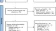
Results: The search identified 53 records. Case reports, letters/notes, and conference abstracts were excluded, resulting in 24 reports assessed by full-text, 9 research articles, 14 reviews, and a book chapter. Five retrospective research studies were found eligible for the review. Detailed MRI features of OCI were only described in two studies of patients with pain where they encompassed manifest subchondral iliac sclerosis often accompanied by bone marrow edema (BME) located peripheral to the sclerosis and displaying a continuous distribution and frequently accompanied by sacral BME. Erosions were rare and ankylosis did not occur. Fat deposition in the bone marrow was frequent and similar to BME often located to anterior strain-related joint areas. The QUADAS-2 assessments revealed risks of bias in all studies analyzed, especially regarding general applicability of the MRI features.
Conclusion: There is a lack of valid data describing characteristic MRI features in general groups of OCI patients with and without pain.
期刊介绍:
Skeletal Radiology provides a forum for the dissemination of current knowledge and information dealing with disorders of the musculoskeletal system including the spine. While emphasizing the radiological aspects of the many varied skeletal abnormalities, the journal also adopts an interdisciplinary approach, reflecting the membership of the International Skeletal Society. Thus, the anatomical, pathological, physiological, clinical, metabolic and epidemiological aspects of the many entities affecting the skeleton receive appropriate consideration.
This is the Journal of the International Skeletal Society and the Official Journal of the Society of Skeletal Radiology and the Australasian Musculoskelelal Imaging Group.


 分享
分享
 求助内容:
求助内容: 应助结果提醒方式:
 扫码关注我们
