Florian Hagen, Linda Vorberg, Florian Thamm, Hendrik Ditt, Andreas Maier, Jan Michael Brendel, Patrick Ghibes, Malte Niklas Bongers, Patrick Krumm, Konstantin Nikolaou, Marius Horger
{"title":"使用基于人工智能的算法改进未增强计算机断层扫描对小肺栓塞的检测--一项单中心回顾性研究。","authors":"Florian Hagen, Linda Vorberg, Florian Thamm, Hendrik Ditt, Andreas Maier, Jan Michael Brendel, Patrick Ghibes, Malte Niklas Bongers, Patrick Krumm, Konstantin Nikolaou, Marius Horger","doi":"10.1007/s10554-024-03222-8","DOIUrl":null,"url":null,"abstract":"<p><p>To preliminarily verify the feasibility of a deep-learning (DL) artificial intelligence (AI) model to localize pulmonary embolism (PE) on unenhanced chest-CT by comparison with pulmonary artery (PA) CT angiography (CTA). In a monocentric study, we retrospectively reviewed 99 oncological patients (median age in years: 64 (range: 28-92 years); percentage of female: 39.4%) who received unenhanced and contrast-enhanced chest CT examinations in one session between January 2020 and October 2022 and who were diagnosed incidentally with PE. Findings in the unenhanced images were correlated with the contrast-enhanced images, which were considered the gold standard for central, segmental and subsegmental PE. The new algorithm was trained and tested based on the 99 unenhanced chest-CT image data sets. Based on them, candidate boxes, which were output by the model, were post-processed by evaluating whether the predicted box intersects with the patient's lung segmentation at any position. The AI-based algorithm proved to have an overall sensitivity of 54.5% for central, of 81.9% for segmental and 80.0% for subsegmental PE if taking n = 20 candidate boxes into account. Depending on the localization of the pulmonary embolism, the detection rate for only one box was: 18.1% central, 34.7% segmental and 0.0% subsegmental. The median volume of the clots differed significantly between the three subgroups and was 846.5 mm<sup>3</sup> (IQR:591.1-964.8) in central, 201.3 mm<sup>3</sup> (IQR:98.3-390.9) in segmental and 110.6 mm<sup>3</sup> (IQR:94.3-128.0) in subsegmental PA (p < 0.05). The new algorithm proved to have high sensitivity in detecting PE in particular in segmental/subsegmental localization and may guide to decide whether a second contrast enhanced CT is necessary.</p>","PeriodicalId":94227,"journal":{"name":"The international journal of cardiovascular imaging","volume":" ","pages":"2293-2304"},"PeriodicalIF":1.5000,"publicationDate":"2024-11-01","publicationTypes":"Journal Article","fieldsOfStudy":null,"isOpenAccess":false,"openAccessPdf":"","citationCount":"0","resultStr":"{\"title\":\"Improved detection of small pulmonary embolism on unenhanced computed tomography using an artificial intelligence-based algorithm - a single centre retrospective study.\",\"authors\":\"Florian Hagen, Linda Vorberg, Florian Thamm, Hendrik Ditt, Andreas Maier, Jan Michael Brendel, Patrick Ghibes, Malte Niklas Bongers, Patrick Krumm, Konstantin Nikolaou, Marius Horger\",\"doi\":\"10.1007/s10554-024-03222-8\",\"DOIUrl\":null,\"url\":null,\"abstract\":\"<p><p>To preliminarily verify the feasibility of a deep-learning (DL) artificial intelligence (AI) model to localize pulmonary embolism (PE) on unenhanced chest-CT by comparison with pulmonary artery (PA) CT angiography (CTA). In a monocentric study, we retrospectively reviewed 99 oncological patients (median age in years: 64 (range: 28-92 years); percentage of female: 39.4%) who received unenhanced and contrast-enhanced chest CT examinations in one session between January 2020 and October 2022 and who were diagnosed incidentally with PE. Findings in the unenhanced images were correlated with the contrast-enhanced images, which were considered the gold standard for central, segmental and subsegmental PE. The new algorithm was trained and tested based on the 99 unenhanced chest-CT image data sets. Based on them, candidate boxes, which were output by the model, were post-processed by evaluating whether the predicted box intersects with the patient's lung segmentation at any position. The AI-based algorithm proved to have an overall sensitivity of 54.5% for central, of 81.9% for segmental and 80.0% for subsegmental PE if taking n = 20 candidate boxes into account. Depending on the localization of the pulmonary embolism, the detection rate for only one box was: 18.1% central, 34.7% segmental and 0.0% subsegmental. The median volume of the clots differed significantly between the three subgroups and was 846.5 mm<sup>3</sup> (IQR:591.1-964.8) in central, 201.3 mm<sup>3</sup> (IQR:98.3-390.9) in segmental and 110.6 mm<sup>3</sup> (IQR:94.3-128.0) in subsegmental PA (p < 0.05). The new algorithm proved to have high sensitivity in detecting PE in particular in segmental/subsegmental localization and may guide to decide whether a second contrast enhanced CT is necessary.</p>\",\"PeriodicalId\":94227,\"journal\":{\"name\":\"The international journal of cardiovascular imaging\",\"volume\":\" \",\"pages\":\"2293-2304\"},\"PeriodicalIF\":1.5000,\"publicationDate\":\"2024-11-01\",\"publicationTypes\":\"Journal Article\",\"fieldsOfStudy\":null,\"isOpenAccess\":false,\"openAccessPdf\":\"\",\"citationCount\":\"0\",\"resultStr\":null,\"platform\":\"Semanticscholar\",\"paperid\":null,\"PeriodicalName\":\"The international journal of cardiovascular imaging\",\"FirstCategoryId\":\"1085\",\"ListUrlMain\":\"https://doi.org/10.1007/s10554-024-03222-8\",\"RegionNum\":0,\"RegionCategory\":null,\"ArticlePicture\":[],\"TitleCN\":null,\"AbstractTextCN\":null,\"PMCID\":null,\"EPubDate\":\"2024/8/28 0:00:00\",\"PubModel\":\"Epub\",\"JCR\":\"\",\"JCRName\":\"\",\"Score\":null,\"Total\":0}","platform":"Semanticscholar","paperid":null,"PeriodicalName":"The international journal of cardiovascular imaging","FirstCategoryId":"1085","ListUrlMain":"https://doi.org/10.1007/s10554-024-03222-8","RegionNum":0,"RegionCategory":null,"ArticlePicture":[],"TitleCN":null,"AbstractTextCN":null,"PMCID":null,"EPubDate":"2024/8/28 0:00:00","PubModel":"Epub","JCR":"","JCRName":"","Score":null,"Total":0}
引用次数: 0
摘要
通过与肺动脉(PA)CT 血管造影术(CTA)比较,初步验证深度学习(DL)人工智能(AI)模型在未增强胸部 CT 上定位肺栓塞(PE)的可行性。在一项单中心研究中,我们回顾性地检查了 99 名肿瘤患者(中位年龄:64 岁(范围:28-92 岁)):中位年龄:64 岁(范围:28-92 岁);女性比例:39.4%),这些患者在 2020 年 1 月至 2022 年 10 月期间接受了一次未增强和对比增强胸部 CT 检查,并被偶然诊断出患有 PE。未增强图像的结果与对比增强图像相关,对比增强图像被认为是中心性、节段性和亚节段性 PE 的金标准。新算法根据 99 个未增强胸部 CT 图像数据集进行了训练和测试。在此基础上,对模型输出的候选方框进行后处理,评估预测方框是否在任何位置与患者肺部分割相交。事实证明,如果考虑到 n = 20 个候选框,基于人工智能的算法对中心型 PE 的总体灵敏度为 54.5%,对节段型 PE 的灵敏度为 81.9%,对亚节段型 PE 的灵敏度为 80.0%。根据肺栓塞的定位情况,仅一个方框的检出率分别为:中心性 18.1%、节段性 34.7% 和亚节段性 0.0%。三个亚组的血块体积中位数差异显著,中央型 PA 为 846.5 立方毫米(IQR:591.1-964.8),节段型 PA 为 201.3 立方毫米(IQR:98.3-390.9),亚节段型 PA 为 110.6 立方毫米(IQR:94.3-128.0)(p
Improved detection of small pulmonary embolism on unenhanced computed tomography using an artificial intelligence-based algorithm - a single centre retrospective study.
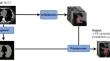
To preliminarily verify the feasibility of a deep-learning (DL) artificial intelligence (AI) model to localize pulmonary embolism (PE) on unenhanced chest-CT by comparison with pulmonary artery (PA) CT angiography (CTA). In a monocentric study, we retrospectively reviewed 99 oncological patients (median age in years: 64 (range: 28-92 years); percentage of female: 39.4%) who received unenhanced and contrast-enhanced chest CT examinations in one session between January 2020 and October 2022 and who were diagnosed incidentally with PE. Findings in the unenhanced images were correlated with the contrast-enhanced images, which were considered the gold standard for central, segmental and subsegmental PE. The new algorithm was trained and tested based on the 99 unenhanced chest-CT image data sets. Based on them, candidate boxes, which were output by the model, were post-processed by evaluating whether the predicted box intersects with the patient's lung segmentation at any position. The AI-based algorithm proved to have an overall sensitivity of 54.5% for central, of 81.9% for segmental and 80.0% for subsegmental PE if taking n = 20 candidate boxes into account. Depending on the localization of the pulmonary embolism, the detection rate for only one box was: 18.1% central, 34.7% segmental and 0.0% subsegmental. The median volume of the clots differed significantly between the three subgroups and was 846.5 mm3 (IQR:591.1-964.8) in central, 201.3 mm3 (IQR:98.3-390.9) in segmental and 110.6 mm3 (IQR:94.3-128.0) in subsegmental PA (p < 0.05). The new algorithm proved to have high sensitivity in detecting PE in particular in segmental/subsegmental localization and may guide to decide whether a second contrast enhanced CT is necessary.


 分享
分享

 求助内容:
求助内容: 应助结果提醒方式:
 扫码关注我们
