Sara Z. Jamal, Blake W. Dieckmann, Gary W. McCollum, John S. Penn, Ashwath Jayagopal, MD Imam Uddin
{"title":"通过缺氧成像预测视网膜分支动脉闭塞的原发性神经元细胞损伤","authors":"Sara Z. Jamal, Blake W. Dieckmann, Gary W. McCollum, John S. Penn, Ashwath Jayagopal, MD Imam Uddin","doi":"10.1111/micc.12883","DOIUrl":null,"url":null,"abstract":"<div>\n \n \n <section>\n \n <h3> Purpose</h3>\n \n <p>To develop a reliable method to generate a mouse model of branch retinal artery occlusion (BRAO) using laser-induced thrombosis of a major artery in the mouse retina. Also, to develop a reliable method to detect retinal hypoxia as predictive biomarker for the risk of neuronal cell damage in BRAO.</p>\n </section>\n \n <section>\n \n <h3> Methods</h3>\n \n <p>A reliable and reproducible model of laser-induced BRAO was developed in mouse retina using Rose Bengal. To characterize retinal hypoxia in BRAO, pimonidazole immunostaining and HYPOX-4 molecular imaging methods were used. Terminal deoxynucleotidyl transferase dUTP nick end labelling (TUNEL) was used to characterize neuronal cell damage in the BRAO retina. Expression of mRNA in retinal tissues from BRAO and age-matched control retinas were analyzed using qRT-PCR.</p>\n </section>\n \n <section>\n \n <h3> Results</h3>\n \n <p>Occlusion of a branch retinal artery near the optic nerve head (ONH) caused a pattern of retinal tissue hypoxia covering about 12.5% of the entire retina. TUNEL-positive cells were localized in all layers in BRAO retinal tissue cross sections. In addition, qRT-PCR data analysis suggests that BRAO is associated with both inflammation and hypoxia.</p>\n </section>\n \n <section>\n \n <h3> Conclusions</h3>\n \n <p>This study provides a reliable method for BRAO in mouse retina and demonstrates the utility of molecular imaging method to detect retinal hypoxia as predictive biomarker for the risk of neuronal cell damage in BRAO. In addition, our data suggest that BRAO retinas are associated with inflammation and also associated with hypoxia-related neuronal cell damage.</p>\n </section>\n \n <section>\n \n <h3> Perspectives</h3>\n \n <p>Imaging areas of retinal hypoxia may provide accurate diagnosis, evaluating retinal tissue injury from BRAO.</p>\n </section>\n </div>","PeriodicalId":18459,"journal":{"name":"Microcirculation","volume":"31 7","pages":""},"PeriodicalIF":2.0000,"publicationDate":"2024-08-30","publicationTypes":"Journal Article","fieldsOfStudy":null,"isOpenAccess":false,"openAccessPdf":"https://onlinelibrary.wiley.com/doi/epdf/10.1111/micc.12883","citationCount":"0","resultStr":"{\"title\":\"Imaging Hypoxia to Predict Primary Neuronal Cell Damage in Branch Retinal Artery Occlusion\",\"authors\":\"Sara Z. Jamal, Blake W. Dieckmann, Gary W. McCollum, John S. Penn, Ashwath Jayagopal, MD Imam Uddin\",\"doi\":\"10.1111/micc.12883\",\"DOIUrl\":null,\"url\":null,\"abstract\":\"<div>\\n \\n \\n <section>\\n \\n <h3> Purpose</h3>\\n \\n <p>To develop a reliable method to generate a mouse model of branch retinal artery occlusion (BRAO) using laser-induced thrombosis of a major artery in the mouse retina. Also, to develop a reliable method to detect retinal hypoxia as predictive biomarker for the risk of neuronal cell damage in BRAO.</p>\\n </section>\\n \\n <section>\\n \\n <h3> Methods</h3>\\n \\n <p>A reliable and reproducible model of laser-induced BRAO was developed in mouse retina using Rose Bengal. To characterize retinal hypoxia in BRAO, pimonidazole immunostaining and HYPOX-4 molecular imaging methods were used. Terminal deoxynucleotidyl transferase dUTP nick end labelling (TUNEL) was used to characterize neuronal cell damage in the BRAO retina. Expression of mRNA in retinal tissues from BRAO and age-matched control retinas were analyzed using qRT-PCR.</p>\\n </section>\\n \\n <section>\\n \\n <h3> Results</h3>\\n \\n <p>Occlusion of a branch retinal artery near the optic nerve head (ONH) caused a pattern of retinal tissue hypoxia covering about 12.5% of the entire retina. TUNEL-positive cells were localized in all layers in BRAO retinal tissue cross sections. In addition, qRT-PCR data analysis suggests that BRAO is associated with both inflammation and hypoxia.</p>\\n </section>\\n \\n <section>\\n \\n <h3> Conclusions</h3>\\n \\n <p>This study provides a reliable method for BRAO in mouse retina and demonstrates the utility of molecular imaging method to detect retinal hypoxia as predictive biomarker for the risk of neuronal cell damage in BRAO. In addition, our data suggest that BRAO retinas are associated with inflammation and also associated with hypoxia-related neuronal cell damage.</p>\\n </section>\\n \\n <section>\\n \\n <h3> Perspectives</h3>\\n \\n <p>Imaging areas of retinal hypoxia may provide accurate diagnosis, evaluating retinal tissue injury from BRAO.</p>\\n </section>\\n </div>\",\"PeriodicalId\":18459,\"journal\":{\"name\":\"Microcirculation\",\"volume\":\"31 7\",\"pages\":\"\"},\"PeriodicalIF\":2.0000,\"publicationDate\":\"2024-08-30\",\"publicationTypes\":\"Journal Article\",\"fieldsOfStudy\":null,\"isOpenAccess\":false,\"openAccessPdf\":\"https://onlinelibrary.wiley.com/doi/epdf/10.1111/micc.12883\",\"citationCount\":\"0\",\"resultStr\":null,\"platform\":\"Semanticscholar\",\"paperid\":null,\"PeriodicalName\":\"Microcirculation\",\"FirstCategoryId\":\"3\",\"ListUrlMain\":\"https://onlinelibrary.wiley.com/doi/10.1111/micc.12883\",\"RegionNum\":4,\"RegionCategory\":\"医学\",\"ArticlePicture\":[],\"TitleCN\":null,\"AbstractTextCN\":null,\"PMCID\":null,\"EPubDate\":\"\",\"PubModel\":\"\",\"JCR\":\"Q3\",\"JCRName\":\"HEMATOLOGY\",\"Score\":null,\"Total\":0}","platform":"Semanticscholar","paperid":null,"PeriodicalName":"Microcirculation","FirstCategoryId":"3","ListUrlMain":"https://onlinelibrary.wiley.com/doi/10.1111/micc.12883","RegionNum":4,"RegionCategory":"医学","ArticlePicture":[],"TitleCN":null,"AbstractTextCN":null,"PMCID":null,"EPubDate":"","PubModel":"","JCR":"Q3","JCRName":"HEMATOLOGY","Score":null,"Total":0}
Imaging Hypoxia to Predict Primary Neuronal Cell Damage in Branch Retinal Artery Occlusion
Purpose
To develop a reliable method to generate a mouse model of branch retinal artery occlusion (BRAO) using laser-induced thrombosis of a major artery in the mouse retina. Also, to develop a reliable method to detect retinal hypoxia as predictive biomarker for the risk of neuronal cell damage in BRAO.
Methods
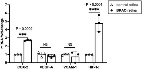
A reliable and reproducible model of laser-induced BRAO was developed in mouse retina using Rose Bengal. To characterize retinal hypoxia in BRAO, pimonidazole immunostaining and HYPOX-4 molecular imaging methods were used. Terminal deoxynucleotidyl transferase dUTP nick end labelling (TUNEL) was used to characterize neuronal cell damage in the BRAO retina. Expression of mRNA in retinal tissues from BRAO and age-matched control retinas were analyzed using qRT-PCR.
Results
Occlusion of a branch retinal artery near the optic nerve head (ONH) caused a pattern of retinal tissue hypoxia covering about 12.5% of the entire retina. TUNEL-positive cells were localized in all layers in BRAO retinal tissue cross sections. In addition, qRT-PCR data analysis suggests that BRAO is associated with both inflammation and hypoxia.
Conclusions
This study provides a reliable method for BRAO in mouse retina and demonstrates the utility of molecular imaging method to detect retinal hypoxia as predictive biomarker for the risk of neuronal cell damage in BRAO. In addition, our data suggest that BRAO retinas are associated with inflammation and also associated with hypoxia-related neuronal cell damage.
Perspectives
Imaging areas of retinal hypoxia may provide accurate diagnosis, evaluating retinal tissue injury from BRAO.
期刊介绍:
The journal features original contributions that are the result of investigations contributing significant new information relating to the vascular and lymphatic microcirculation addressed at the intact animal, organ, cellular, or molecular level. Papers describe applications of the methods of physiology, biophysics, bioengineering, genetics, cell biology, biochemistry, and molecular biology to problems in microcirculation.
Microcirculation also publishes state-of-the-art reviews that address frontier areas or new advances in technology in the fields of microcirculatory disease and function. Specific areas of interest include: Angiogenesis, growth and remodeling; Transport and exchange of gasses and solutes; Rheology and biorheology; Endothelial cell biology and metabolism; Interactions between endothelium, smooth muscle, parenchymal cells, leukocytes and platelets; Regulation of vasomotor tone; and Microvascular structures, imaging and morphometry. Papers also describe innovations in experimental techniques and instrumentation for studying all aspects of microcirculatory structure and function.


 分享
分享
 求助内容:
求助内容: 应助结果提醒方式:
 扫码关注我们
