Vincent-Béni Sèna Zossou, Freddy Houéhanou Rodrigue Gnangnon, Olivier Biaou, Florent de Vathaire, Rodrigue S Allodji, Eugène C Ezin
{"title":"基于计算机断层扫描图像的肝细胞癌和转移瘤自动诊断。","authors":"Vincent-Béni Sèna Zossou, Freddy Houéhanou Rodrigue Gnangnon, Olivier Biaou, Florent de Vathaire, Rodrigue S Allodji, Eugène C Ezin","doi":"10.1007/s10278-024-01192-w","DOIUrl":null,"url":null,"abstract":"<p><p>Liver cancer, a leading cause of cancer mortality, is often diagnosed by analyzing the grayscale variations in liver tissue across different computed tomography (CT) images. However, the intensity similarity can be strong, making it difficult for radiologists to visually identify hepatocellular carcinoma (HCC) and metastases. It is crucial for the management and prevention strategies to accurately differentiate between these two liver cancers. This study proposes an automated system using a convolutional neural network (CNN) to enhance diagnostic accuracy to detect HCC, metastasis, and healthy liver tissue. This system incorporates automatic segmentation and classification. The liver lesions segmentation model is implemented using residual attention U-Net. A 9-layer CNN classifier implements the lesions classification model. Its input is the combination of the results of the segmentation model with original images. The dataset included 300 patients, with 223 used to develop the segmentation model and 77 to test it. These 77 patients also served as inputs for the classification model, consisting of 20 HCC cases, 27 with metastasis, and 30 healthy. The system achieved a mean Dice score of <math><mrow><mn>87.65</mn> <mo>%</mo></mrow> </math> in segmentation and a mean accuracy of <math><mrow><mn>93.97</mn> <mo>%</mo></mrow> </math> in classification, both in the test phase. The proposed method is a preliminary study with great potential in helping radiologists diagnose liver cancers.</p>","PeriodicalId":516858,"journal":{"name":"Journal of imaging informatics in medicine","volume":" ","pages":"873-886"},"PeriodicalIF":0.0000,"publicationDate":"2025-04-01","publicationTypes":"Journal Article","fieldsOfStudy":null,"isOpenAccess":false,"openAccessPdf":"https://www.ncbi.nlm.nih.gov/pmc/articles/PMC11950545/pdf/","citationCount":"0","resultStr":"{\"title\":\"Automatic Diagnosis of Hepatocellular Carcinoma and Metastases Based on Computed Tomography Images.\",\"authors\":\"Vincent-Béni Sèna Zossou, Freddy Houéhanou Rodrigue Gnangnon, Olivier Biaou, Florent de Vathaire, Rodrigue S Allodji, Eugène C Ezin\",\"doi\":\"10.1007/s10278-024-01192-w\",\"DOIUrl\":null,\"url\":null,\"abstract\":\"<p><p>Liver cancer, a leading cause of cancer mortality, is often diagnosed by analyzing the grayscale variations in liver tissue across different computed tomography (CT) images. However, the intensity similarity can be strong, making it difficult for radiologists to visually identify hepatocellular carcinoma (HCC) and metastases. It is crucial for the management and prevention strategies to accurately differentiate between these two liver cancers. This study proposes an automated system using a convolutional neural network (CNN) to enhance diagnostic accuracy to detect HCC, metastasis, and healthy liver tissue. This system incorporates automatic segmentation and classification. The liver lesions segmentation model is implemented using residual attention U-Net. A 9-layer CNN classifier implements the lesions classification model. Its input is the combination of the results of the segmentation model with original images. The dataset included 300 patients, with 223 used to develop the segmentation model and 77 to test it. These 77 patients also served as inputs for the classification model, consisting of 20 HCC cases, 27 with metastasis, and 30 healthy. The system achieved a mean Dice score of <math><mrow><mn>87.65</mn> <mo>%</mo></mrow> </math> in segmentation and a mean accuracy of <math><mrow><mn>93.97</mn> <mo>%</mo></mrow> </math> in classification, both in the test phase. The proposed method is a preliminary study with great potential in helping radiologists diagnose liver cancers.</p>\",\"PeriodicalId\":516858,\"journal\":{\"name\":\"Journal of imaging informatics in medicine\",\"volume\":\" \",\"pages\":\"873-886\"},\"PeriodicalIF\":0.0000,\"publicationDate\":\"2025-04-01\",\"publicationTypes\":\"Journal Article\",\"fieldsOfStudy\":null,\"isOpenAccess\":false,\"openAccessPdf\":\"https://www.ncbi.nlm.nih.gov/pmc/articles/PMC11950545/pdf/\",\"citationCount\":\"0\",\"resultStr\":null,\"platform\":\"Semanticscholar\",\"paperid\":null,\"PeriodicalName\":\"Journal of imaging informatics in medicine\",\"FirstCategoryId\":\"1085\",\"ListUrlMain\":\"https://doi.org/10.1007/s10278-024-01192-w\",\"RegionNum\":0,\"RegionCategory\":null,\"ArticlePicture\":[],\"TitleCN\":null,\"AbstractTextCN\":null,\"PMCID\":null,\"EPubDate\":\"2024/9/3 0:00:00\",\"PubModel\":\"Epub\",\"JCR\":\"\",\"JCRName\":\"\",\"Score\":null,\"Total\":0}","platform":"Semanticscholar","paperid":null,"PeriodicalName":"Journal of imaging informatics in medicine","FirstCategoryId":"1085","ListUrlMain":"https://doi.org/10.1007/s10278-024-01192-w","RegionNum":0,"RegionCategory":null,"ArticlePicture":[],"TitleCN":null,"AbstractTextCN":null,"PMCID":null,"EPubDate":"2024/9/3 0:00:00","PubModel":"Epub","JCR":"","JCRName":"","Score":null,"Total":0}
Automatic Diagnosis of Hepatocellular Carcinoma and Metastases Based on Computed Tomography Images.
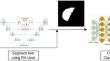
Liver cancer, a leading cause of cancer mortality, is often diagnosed by analyzing the grayscale variations in liver tissue across different computed tomography (CT) images. However, the intensity similarity can be strong, making it difficult for radiologists to visually identify hepatocellular carcinoma (HCC) and metastases. It is crucial for the management and prevention strategies to accurately differentiate between these two liver cancers. This study proposes an automated system using a convolutional neural network (CNN) to enhance diagnostic accuracy to detect HCC, metastasis, and healthy liver tissue. This system incorporates automatic segmentation and classification. The liver lesions segmentation model is implemented using residual attention U-Net. A 9-layer CNN classifier implements the lesions classification model. Its input is the combination of the results of the segmentation model with original images. The dataset included 300 patients, with 223 used to develop the segmentation model and 77 to test it. These 77 patients also served as inputs for the classification model, consisting of 20 HCC cases, 27 with metastasis, and 30 healthy. The system achieved a mean Dice score of in segmentation and a mean accuracy of in classification, both in the test phase. The proposed method is a preliminary study with great potential in helping radiologists diagnose liver cancers.


 分享
分享

 求助内容:
求助内容: 应助结果提醒方式:
 扫码关注我们
