{"title":"利用 CD3+CD4+CD26- T 细胞对典型霍奇金淋巴瘤进行隐性流式细胞术诊断","authors":"Curtis Gravenmier, Jinming Song, Haipeng Shao","doi":"10.1002/jcla.25096","DOIUrl":null,"url":null,"abstract":"<div>\n \n \n <section>\n \n <h3> Background</h3>\n \n <p>Flow cytometry is not routinely performed in clinical laboratories for the diagnosis of classic Hodgkin lymphoma (CHL).</p>\n </section>\n \n <section>\n \n <h3> Methods</h3>\n \n <p>Fourteen cases of CHL and 132 cases of the control group were studied by 10-color flow cytometry, with markers including CD3, CD4, CD7, CD8, and CD26, as well as calculated parameters such as the CD4:CD8 ratio, percent CD3<sup>+</sup>CD4<sup>+</sup>CD26<sup>−</sup> T-cells of CD3<sup>+</sup>CD4<sup>+</sup> T-cells, percent CD3<sup>+</sup>CD4<sup>+</sup>CD26<sup>−</sup> T-cells of total events, CD7 coefficient of variation among CD3<sup>+</sup>CD4<sup>+</sup>CD26<sup>−</sup> T-cells, and CD7 median fluorescence intensity of CD3<sup>+</sup>CD4<sup>+</sup>CD26<sup>−</sup> T-cells relative to CD3<sup>+</sup>CD8<sup>+</sup> T-cells.</p>\n </section>\n \n <section>\n \n <h3> Results</h3>\n \n <p>CHL cases showed a median percent CD3<sup>+</sup>CD4<sup>+</sup>CD26<sup>−</sup> of CD3<sup>+</sup>CD4<sup>+</sup> T-cells of 72.3% with range from 41.1% to 94.4%, median percent CD3<sup>+</sup>CD4<sup>+</sup>CD26<sup>−</sup> T-cells of total events of 17.4% with range from 4.6% to 52.5%, CD7 coefficient of variation among CD3<sup>+</sup>CD4<sup>+</sup>CD26<sup>−</sup> T-cells less than 100%, and CD7 median fluorescence intensity of CD3<sup>+</sup>CD4<sup>+</sup>CD26<sup>−</sup> T-cells relative to CD3<sup>+</sup>CD8<sup>+</sup> T-cells of 1.7 with range from 0.4 to 3.5. In the control group, every entity showed some degree of overlap with CHL in terms of these parameters. A “Hodgkin score” was thus constructed to enhance separation of CHL from other entities. A threshold Hodgkin score of 15.35 achieved a sensitivity of 78.6% and specificity of 96.2% in the diagnosis of CHL. Incorporating the Hodgkin score into a simple algorithm raises the specificity to 100%.</p>\n </section>\n \n <section>\n \n <h3> Conclusion</h3>\n \n <p>In this study, we used flow cytometry to demonstrate increased CD3<sup>+</sup>CD4<sup>+</sup>CD26<sup>−</sup> T-cells in CHL, and derived a Hodgkin score for the diagnosis of CHL.</p>\n </section>\n </div>","PeriodicalId":15509,"journal":{"name":"Journal of Clinical Laboratory Analysis","volume":"38 17-18","pages":""},"PeriodicalIF":2.6000,"publicationDate":"2024-09-05","publicationTypes":"Journal Article","fieldsOfStudy":null,"isOpenAccess":false,"openAccessPdf":"https://onlinelibrary.wiley.com/doi/epdf/10.1002/jcla.25096","citationCount":"0","resultStr":"{\"title\":\"Implicit Flow Cytometric Diagnosis of Classic Hodgkin Lymphoma Using CD3+CD4+CD26− T-Cells\",\"authors\":\"Curtis Gravenmier, Jinming Song, Haipeng Shao\",\"doi\":\"10.1002/jcla.25096\",\"DOIUrl\":null,\"url\":null,\"abstract\":\"<div>\\n \\n \\n <section>\\n \\n <h3> Background</h3>\\n \\n <p>Flow cytometry is not routinely performed in clinical laboratories for the diagnosis of classic Hodgkin lymphoma (CHL).</p>\\n </section>\\n \\n <section>\\n \\n <h3> Methods</h3>\\n \\n <p>Fourteen cases of CHL and 132 cases of the control group were studied by 10-color flow cytometry, with markers including CD3, CD4, CD7, CD8, and CD26, as well as calculated parameters such as the CD4:CD8 ratio, percent CD3<sup>+</sup>CD4<sup>+</sup>CD26<sup>−</sup> T-cells of CD3<sup>+</sup>CD4<sup>+</sup> T-cells, percent CD3<sup>+</sup>CD4<sup>+</sup>CD26<sup>−</sup> T-cells of total events, CD7 coefficient of variation among CD3<sup>+</sup>CD4<sup>+</sup>CD26<sup>−</sup> T-cells, and CD7 median fluorescence intensity of CD3<sup>+</sup>CD4<sup>+</sup>CD26<sup>−</sup> T-cells relative to CD3<sup>+</sup>CD8<sup>+</sup> T-cells.</p>\\n </section>\\n \\n <section>\\n \\n <h3> Results</h3>\\n \\n <p>CHL cases showed a median percent CD3<sup>+</sup>CD4<sup>+</sup>CD26<sup>−</sup> of CD3<sup>+</sup>CD4<sup>+</sup> T-cells of 72.3% with range from 41.1% to 94.4%, median percent CD3<sup>+</sup>CD4<sup>+</sup>CD26<sup>−</sup> T-cells of total events of 17.4% with range from 4.6% to 52.5%, CD7 coefficient of variation among CD3<sup>+</sup>CD4<sup>+</sup>CD26<sup>−</sup> T-cells less than 100%, and CD7 median fluorescence intensity of CD3<sup>+</sup>CD4<sup>+</sup>CD26<sup>−</sup> T-cells relative to CD3<sup>+</sup>CD8<sup>+</sup> T-cells of 1.7 with range from 0.4 to 3.5. In the control group, every entity showed some degree of overlap with CHL in terms of these parameters. A “Hodgkin score” was thus constructed to enhance separation of CHL from other entities. A threshold Hodgkin score of 15.35 achieved a sensitivity of 78.6% and specificity of 96.2% in the diagnosis of CHL. Incorporating the Hodgkin score into a simple algorithm raises the specificity to 100%.</p>\\n </section>\\n \\n <section>\\n \\n <h3> Conclusion</h3>\\n \\n <p>In this study, we used flow cytometry to demonstrate increased CD3<sup>+</sup>CD4<sup>+</sup>CD26<sup>−</sup> T-cells in CHL, and derived a Hodgkin score for the diagnosis of CHL.</p>\\n </section>\\n </div>\",\"PeriodicalId\":15509,\"journal\":{\"name\":\"Journal of Clinical Laboratory Analysis\",\"volume\":\"38 17-18\",\"pages\":\"\"},\"PeriodicalIF\":2.6000,\"publicationDate\":\"2024-09-05\",\"publicationTypes\":\"Journal Article\",\"fieldsOfStudy\":null,\"isOpenAccess\":false,\"openAccessPdf\":\"https://onlinelibrary.wiley.com/doi/epdf/10.1002/jcla.25096\",\"citationCount\":\"0\",\"resultStr\":null,\"platform\":\"Semanticscholar\",\"paperid\":null,\"PeriodicalName\":\"Journal of Clinical Laboratory Analysis\",\"FirstCategoryId\":\"3\",\"ListUrlMain\":\"https://onlinelibrary.wiley.com/doi/10.1002/jcla.25096\",\"RegionNum\":4,\"RegionCategory\":\"医学\",\"ArticlePicture\":[],\"TitleCN\":null,\"AbstractTextCN\":null,\"PMCID\":null,\"EPubDate\":\"\",\"PubModel\":\"\",\"JCR\":\"Q2\",\"JCRName\":\"MEDICAL LABORATORY TECHNOLOGY\",\"Score\":null,\"Total\":0}","platform":"Semanticscholar","paperid":null,"PeriodicalName":"Journal of Clinical Laboratory Analysis","FirstCategoryId":"3","ListUrlMain":"https://onlinelibrary.wiley.com/doi/10.1002/jcla.25096","RegionNum":4,"RegionCategory":"医学","ArticlePicture":[],"TitleCN":null,"AbstractTextCN":null,"PMCID":null,"EPubDate":"","PubModel":"","JCR":"Q2","JCRName":"MEDICAL LABORATORY TECHNOLOGY","Score":null,"Total":0}
Implicit Flow Cytometric Diagnosis of Classic Hodgkin Lymphoma Using CD3+CD4+CD26− T-Cells
Background
Flow cytometry is not routinely performed in clinical laboratories for the diagnosis of classic Hodgkin lymphoma (CHL).
Methods
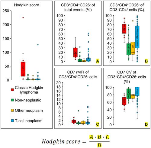
Fourteen cases of CHL and 132 cases of the control group were studied by 10-color flow cytometry, with markers including CD3, CD4, CD7, CD8, and CD26, as well as calculated parameters such as the CD4:CD8 ratio, percent CD3+CD4+CD26− T-cells of CD3+CD4+ T-cells, percent CD3+CD4+CD26− T-cells of total events, CD7 coefficient of variation among CD3+CD4+CD26− T-cells, and CD7 median fluorescence intensity of CD3+CD4+CD26− T-cells relative to CD3+CD8+ T-cells.
Results
CHL cases showed a median percent CD3+CD4+CD26− of CD3+CD4+ T-cells of 72.3% with range from 41.1% to 94.4%, median percent CD3+CD4+CD26− T-cells of total events of 17.4% with range from 4.6% to 52.5%, CD7 coefficient of variation among CD3+CD4+CD26− T-cells less than 100%, and CD7 median fluorescence intensity of CD3+CD4+CD26− T-cells relative to CD3+CD8+ T-cells of 1.7 with range from 0.4 to 3.5. In the control group, every entity showed some degree of overlap with CHL in terms of these parameters. A “Hodgkin score” was thus constructed to enhance separation of CHL from other entities. A threshold Hodgkin score of 15.35 achieved a sensitivity of 78.6% and specificity of 96.2% in the diagnosis of CHL. Incorporating the Hodgkin score into a simple algorithm raises the specificity to 100%.
Conclusion
In this study, we used flow cytometry to demonstrate increased CD3+CD4+CD26− T-cells in CHL, and derived a Hodgkin score for the diagnosis of CHL.
期刊介绍:
Journal of Clinical Laboratory Analysis publishes original articles on newly developing modes of technology and laboratory assays, with emphasis on their application in current and future clinical laboratory testing. This includes reports from the following fields: immunochemistry and toxicology, hematology and hematopathology, immunopathology, molecular diagnostics, microbiology, genetic testing, immunohematology, and clinical chemistry.


 分享
分享
 求助内容:
求助内容: 应助结果提醒方式:
 扫码关注我们
