Sónia Castanheira, David López-Escarpa, Alberto Paradela, Francisco García-Del Portillo
{"title":"体内交联揭示了沙门氏菌分裂体中 PBP3 和 PBP3SAL 竞相占据的情况。","authors":"Sónia Castanheira, David López-Escarpa, Alberto Paradela, Francisco García-Del Portillo","doi":"10.1111/mmi.15309","DOIUrl":null,"url":null,"abstract":"<p><p>Bacterial cell division is orchestrated by proteins that assemble in dynamic complexes collectively known as the divisome. Essential monofunctional enzymes with glycosyltransferase or transpeptidase (TPase) activities, FtsW and FtsI respectively, engage in the synthesis of septal peptidoglycan (sPG). Enigmatically, Salmonella has two TPases that can promote cell division independently: FtsI (PBP3) and the pathogen-specific paralogue PBP3<sub>SAL</sub>. How Salmonella regulates the assembly of the sPG synthase complex with these two TPases, is unknown. Here, we characterized Salmonella division complexes in wild-type cells and isogenic mutants lacking PBP3 or PBP3<sub>SAL</sub>. The complexes were cross-linked in vivo and pulled down with antibodies recognizing each enzyme. Proteomics of the immunoprecipitates showed that PBP3 and PBP3<sub>SAL</sub> do not extensively cross-link in wild type cells, supporting the presence of independent complexes. More than 40 proteins cross-link in complexes in which these two TPases are present. Those identified with high scores include FtsA, FtsK, FtsQLB, FtsW, PBP1B, SPOR domain-containing proteins (FtsN, DedD, RlpA, DamX), amidase activators (FtsX, EnvC, NlpD) and Tol-Pal proteins. Other cross-linked proteins are the protease Prc, the elongasome TPase PBP2 and, D,D-endo- and D,D-carboxypeptidases. PBP3 and PBP3<sub>SAL</sub> localize at midcell and compete for occupying the division complex in response to environmental cues. Thus, a catalytic-dead PBP3<sub>SAL</sub>-S300A variant impairs cell division in a high osmolarity and acidic condition in which it is produced at levels exceeding those of PBP3. Salmonella may therefore exploit an 'adjustable' divisome to exchange TPases for ensuring cell division in distinct environments and, in this manner, expand its colonization capacities.</p>","PeriodicalId":19006,"journal":{"name":"Molecular Microbiology","volume":" ","pages":"797-818"},"PeriodicalIF":2.6000,"publicationDate":"2024-11-01","publicationTypes":"Journal Article","fieldsOfStudy":null,"isOpenAccess":false,"openAccessPdf":"https://www.ncbi.nlm.nih.gov/pmc/articles/PMC11586514/pdf/","citationCount":"0","resultStr":"{\"title\":\"In Vivo Cross-Linking Sheds Light on the Salmonella Divisome in Which PBP3 and PBP3<sub>SAL</sub> Compete for Occupancy.\",\"authors\":\"Sónia Castanheira, David López-Escarpa, Alberto Paradela, Francisco García-Del Portillo\",\"doi\":\"10.1111/mmi.15309\",\"DOIUrl\":null,\"url\":null,\"abstract\":\"<p><p>Bacterial cell division is orchestrated by proteins that assemble in dynamic complexes collectively known as the divisome. Essential monofunctional enzymes with glycosyltransferase or transpeptidase (TPase) activities, FtsW and FtsI respectively, engage in the synthesis of septal peptidoglycan (sPG). Enigmatically, Salmonella has two TPases that can promote cell division independently: FtsI (PBP3) and the pathogen-specific paralogue PBP3<sub>SAL</sub>. How Salmonella regulates the assembly of the sPG synthase complex with these two TPases, is unknown. Here, we characterized Salmonella division complexes in wild-type cells and isogenic mutants lacking PBP3 or PBP3<sub>SAL</sub>. The complexes were cross-linked in vivo and pulled down with antibodies recognizing each enzyme. Proteomics of the immunoprecipitates showed that PBP3 and PBP3<sub>SAL</sub> do not extensively cross-link in wild type cells, supporting the presence of independent complexes. More than 40 proteins cross-link in complexes in which these two TPases are present. Those identified with high scores include FtsA, FtsK, FtsQLB, FtsW, PBP1B, SPOR domain-containing proteins (FtsN, DedD, RlpA, DamX), amidase activators (FtsX, EnvC, NlpD) and Tol-Pal proteins. Other cross-linked proteins are the protease Prc, the elongasome TPase PBP2 and, D,D-endo- and D,D-carboxypeptidases. PBP3 and PBP3<sub>SAL</sub> localize at midcell and compete for occupying the division complex in response to environmental cues. Thus, a catalytic-dead PBP3<sub>SAL</sub>-S300A variant impairs cell division in a high osmolarity and acidic condition in which it is produced at levels exceeding those of PBP3. Salmonella may therefore exploit an 'adjustable' divisome to exchange TPases for ensuring cell division in distinct environments and, in this manner, expand its colonization capacities.</p>\",\"PeriodicalId\":19006,\"journal\":{\"name\":\"Molecular Microbiology\",\"volume\":\" \",\"pages\":\"797-818\"},\"PeriodicalIF\":2.6000,\"publicationDate\":\"2024-11-01\",\"publicationTypes\":\"Journal Article\",\"fieldsOfStudy\":null,\"isOpenAccess\":false,\"openAccessPdf\":\"https://www.ncbi.nlm.nih.gov/pmc/articles/PMC11586514/pdf/\",\"citationCount\":\"0\",\"resultStr\":null,\"platform\":\"Semanticscholar\",\"paperid\":null,\"PeriodicalName\":\"Molecular Microbiology\",\"FirstCategoryId\":\"99\",\"ListUrlMain\":\"https://doi.org/10.1111/mmi.15309\",\"RegionNum\":2,\"RegionCategory\":\"生物学\",\"ArticlePicture\":[],\"TitleCN\":null,\"AbstractTextCN\":null,\"PMCID\":null,\"EPubDate\":\"2024/9/4 0:00:00\",\"PubModel\":\"Epub\",\"JCR\":\"Q3\",\"JCRName\":\"BIOCHEMISTRY & MOLECULAR BIOLOGY\",\"Score\":null,\"Total\":0}","platform":"Semanticscholar","paperid":null,"PeriodicalName":"Molecular Microbiology","FirstCategoryId":"99","ListUrlMain":"https://doi.org/10.1111/mmi.15309","RegionNum":2,"RegionCategory":"生物学","ArticlePicture":[],"TitleCN":null,"AbstractTextCN":null,"PMCID":null,"EPubDate":"2024/9/4 0:00:00","PubModel":"Epub","JCR":"Q3","JCRName":"BIOCHEMISTRY & MOLECULAR BIOLOGY","Score":null,"Total":0}
In Vivo Cross-Linking Sheds Light on the Salmonella Divisome in Which PBP3 and PBP3SAL Compete for Occupancy.
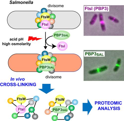
Bacterial cell division is orchestrated by proteins that assemble in dynamic complexes collectively known as the divisome. Essential monofunctional enzymes with glycosyltransferase or transpeptidase (TPase) activities, FtsW and FtsI respectively, engage in the synthesis of septal peptidoglycan (sPG). Enigmatically, Salmonella has two TPases that can promote cell division independently: FtsI (PBP3) and the pathogen-specific paralogue PBP3SAL. How Salmonella regulates the assembly of the sPG synthase complex with these two TPases, is unknown. Here, we characterized Salmonella division complexes in wild-type cells and isogenic mutants lacking PBP3 or PBP3SAL. The complexes were cross-linked in vivo and pulled down with antibodies recognizing each enzyme. Proteomics of the immunoprecipitates showed that PBP3 and PBP3SAL do not extensively cross-link in wild type cells, supporting the presence of independent complexes. More than 40 proteins cross-link in complexes in which these two TPases are present. Those identified with high scores include FtsA, FtsK, FtsQLB, FtsW, PBP1B, SPOR domain-containing proteins (FtsN, DedD, RlpA, DamX), amidase activators (FtsX, EnvC, NlpD) and Tol-Pal proteins. Other cross-linked proteins are the protease Prc, the elongasome TPase PBP2 and, D,D-endo- and D,D-carboxypeptidases. PBP3 and PBP3SAL localize at midcell and compete for occupying the division complex in response to environmental cues. Thus, a catalytic-dead PBP3SAL-S300A variant impairs cell division in a high osmolarity and acidic condition in which it is produced at levels exceeding those of PBP3. Salmonella may therefore exploit an 'adjustable' divisome to exchange TPases for ensuring cell division in distinct environments and, in this manner, expand its colonization capacities.
期刊介绍:
Molecular Microbiology, the leading primary journal in the microbial sciences, publishes molecular studies of Bacteria, Archaea, eukaryotic microorganisms, and their viruses.
Research papers should lead to a deeper understanding of the molecular principles underlying basic physiological processes or mechanisms. Appropriate topics include gene expression and regulation, pathogenicity and virulence, physiology and metabolism, synthesis of macromolecules (proteins, nucleic acids, lipids, polysaccharides, etc), cell biology and subcellular organization, membrane biogenesis and function, traffic and transport, cell-cell communication and signalling pathways, evolution and gene transfer. Articles focused on host responses (cellular or immunological) to pathogens or on microbial ecology should be directed to our sister journals Cellular Microbiology and Environmental Microbiology, respectively.


 分享
分享
 求助内容:
求助内容: 应助结果提醒方式:
 扫码关注我们
