Joeri Kok , Melissa S.A.M. Bevers , Bert van Rietbergen , Edwin H.G. Oei , Ronald Booij
{"title":"在不同辐射剂量下使用光子计数 CT 对骨微观结构进行量化:与 µCT 的比较。","authors":"Joeri Kok , Melissa S.A.M. Bevers , Bert van Rietbergen , Edwin H.G. Oei , Ronald Booij","doi":"10.1016/j.ejrad.2024.111717","DOIUrl":null,"url":null,"abstract":"<div><h3>Purpose</h3><p>Accurate measurements of trabecular bone microarchitecture are required for a proper assessment of bone fragility. Photon-counting detector CT (PCD-CT) has different technical properties than conventional CT, resulting in higher resolution and thereby potentially enabling <em>in-vivo</em> measurement of trabecular microarchitecture. The purpose of this study was to quantify trabecular bone microarchitectural parameters with PCD-CT at varying radiation doses and compare this to µCT as gold standard.</p></div><div><h3>Method</h3><p>Both distal radii, distal tibiae, femoral heads, and two vertebrae were dissected from one human. All specimens were scanned <em>ex-vivo</em> on a PCD-CT system (slice increment 0.1 mm; pixel size 0.1042–0.127 mm) and a µCT system (isotropic voxel size 49–68.4 µm). The radiation doses of the PCD-CT scans were varied from 2.5 to 120 mGy based on the volume CT dose index (CTDI<sub>vol32</sub>). For the PCD-CT scans, contrast-to-noise ratio and trabecular sharpness were calculated and compared between radiation doses. µCT and PCD-CT scans were registered. The trabecular bone was then segmented from all PCD-CT and µCT scans and split into cubes with 6-mm edge length. For each cube, bone volume over total volume, trabecular thickness, trabecular number, and trabecular heterogeneity were calculated and compared between corresponding PCD-CT and µCT cubes.</p></div><div><h3>Results</h3><p>With increasing dose, contrast-to-noise ratio and trabecular sharpness values increased for the PCD-CT images. Already at the lowest dose, high correlations between the trabecular microarchitectural parameters between µCT and PCD-CT were found (R<sup>2</sup> = 0.55–0.95), which improved with increasing radiation dose (R<sup>2</sup> = 0.76–0.96 at 20 mGy).</p></div><div><h3>Conclusions</h3><p>PCD-CT can be used to quantify trabecular bone microarchitecture, with accuracy comparable to µCT and at clinically relevant radiation doses.</p></div>","PeriodicalId":12063,"journal":{"name":"European Journal of Radiology","volume":"181 ","pages":"Article 111717"},"PeriodicalIF":3.3000,"publicationDate":"2024-09-02","publicationTypes":"Journal Article","fieldsOfStudy":null,"isOpenAccess":false,"openAccessPdf":"https://www.sciencedirect.com/science/article/pii/S0720048X24004339/pdfft?md5=707c0b792b17a6ee84bf01abcc85f796&pid=1-s2.0-S0720048X24004339-main.pdf","citationCount":"0","resultStr":"{\"title\":\"Quantification of bone microarchitecture using photon-counting CT at different radiation doses: A comparison with µCT\",\"authors\":\"Joeri Kok , Melissa S.A.M. Bevers , Bert van Rietbergen , Edwin H.G. Oei , Ronald Booij\",\"doi\":\"10.1016/j.ejrad.2024.111717\",\"DOIUrl\":null,\"url\":null,\"abstract\":\"<div><h3>Purpose</h3><p>Accurate measurements of trabecular bone microarchitecture are required for a proper assessment of bone fragility. Photon-counting detector CT (PCD-CT) has different technical properties than conventional CT, resulting in higher resolution and thereby potentially enabling <em>in-vivo</em> measurement of trabecular microarchitecture. The purpose of this study was to quantify trabecular bone microarchitectural parameters with PCD-CT at varying radiation doses and compare this to µCT as gold standard.</p></div><div><h3>Method</h3><p>Both distal radii, distal tibiae, femoral heads, and two vertebrae were dissected from one human. All specimens were scanned <em>ex-vivo</em> on a PCD-CT system (slice increment 0.1 mm; pixel size 0.1042–0.127 mm) and a µCT system (isotropic voxel size 49–68.4 µm). The radiation doses of the PCD-CT scans were varied from 2.5 to 120 mGy based on the volume CT dose index (CTDI<sub>vol32</sub>). For the PCD-CT scans, contrast-to-noise ratio and trabecular sharpness were calculated and compared between radiation doses. µCT and PCD-CT scans were registered. The trabecular bone was then segmented from all PCD-CT and µCT scans and split into cubes with 6-mm edge length. For each cube, bone volume over total volume, trabecular thickness, trabecular number, and trabecular heterogeneity were calculated and compared between corresponding PCD-CT and µCT cubes.</p></div><div><h3>Results</h3><p>With increasing dose, contrast-to-noise ratio and trabecular sharpness values increased for the PCD-CT images. Already at the lowest dose, high correlations between the trabecular microarchitectural parameters between µCT and PCD-CT were found (R<sup>2</sup> = 0.55–0.95), which improved with increasing radiation dose (R<sup>2</sup> = 0.76–0.96 at 20 mGy).</p></div><div><h3>Conclusions</h3><p>PCD-CT can be used to quantify trabecular bone microarchitecture, with accuracy comparable to µCT and at clinically relevant radiation doses.</p></div>\",\"PeriodicalId\":12063,\"journal\":{\"name\":\"European Journal of Radiology\",\"volume\":\"181 \",\"pages\":\"Article 111717\"},\"PeriodicalIF\":3.3000,\"publicationDate\":\"2024-09-02\",\"publicationTypes\":\"Journal Article\",\"fieldsOfStudy\":null,\"isOpenAccess\":false,\"openAccessPdf\":\"https://www.sciencedirect.com/science/article/pii/S0720048X24004339/pdfft?md5=707c0b792b17a6ee84bf01abcc85f796&pid=1-s2.0-S0720048X24004339-main.pdf\",\"citationCount\":\"0\",\"resultStr\":null,\"platform\":\"Semanticscholar\",\"paperid\":null,\"PeriodicalName\":\"European Journal of Radiology\",\"FirstCategoryId\":\"3\",\"ListUrlMain\":\"https://www.sciencedirect.com/science/article/pii/S0720048X24004339\",\"RegionNum\":3,\"RegionCategory\":\"医学\",\"ArticlePicture\":[],\"TitleCN\":null,\"AbstractTextCN\":null,\"PMCID\":null,\"EPubDate\":\"\",\"PubModel\":\"\",\"JCR\":\"Q1\",\"JCRName\":\"RADIOLOGY, NUCLEAR MEDICINE & MEDICAL IMAGING\",\"Score\":null,\"Total\":0}","platform":"Semanticscholar","paperid":null,"PeriodicalName":"European Journal of Radiology","FirstCategoryId":"3","ListUrlMain":"https://www.sciencedirect.com/science/article/pii/S0720048X24004339","RegionNum":3,"RegionCategory":"医学","ArticlePicture":[],"TitleCN":null,"AbstractTextCN":null,"PMCID":null,"EPubDate":"","PubModel":"","JCR":"Q1","JCRName":"RADIOLOGY, NUCLEAR MEDICINE & MEDICAL IMAGING","Score":null,"Total":0}
Quantification of bone microarchitecture using photon-counting CT at different radiation doses: A comparison with µCT
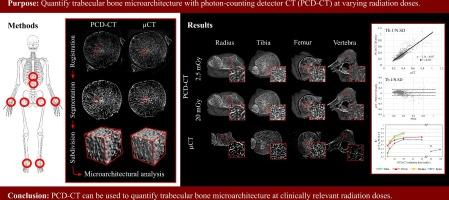
Purpose
Accurate measurements of trabecular bone microarchitecture are required for a proper assessment of bone fragility. Photon-counting detector CT (PCD-CT) has different technical properties than conventional CT, resulting in higher resolution and thereby potentially enabling in-vivo measurement of trabecular microarchitecture. The purpose of this study was to quantify trabecular bone microarchitectural parameters with PCD-CT at varying radiation doses and compare this to µCT as gold standard.
Method
Both distal radii, distal tibiae, femoral heads, and two vertebrae were dissected from one human. All specimens were scanned ex-vivo on a PCD-CT system (slice increment 0.1 mm; pixel size 0.1042–0.127 mm) and a µCT system (isotropic voxel size 49–68.4 µm). The radiation doses of the PCD-CT scans were varied from 2.5 to 120 mGy based on the volume CT dose index (CTDIvol32). For the PCD-CT scans, contrast-to-noise ratio and trabecular sharpness were calculated and compared between radiation doses. µCT and PCD-CT scans were registered. The trabecular bone was then segmented from all PCD-CT and µCT scans and split into cubes with 6-mm edge length. For each cube, bone volume over total volume, trabecular thickness, trabecular number, and trabecular heterogeneity were calculated and compared between corresponding PCD-CT and µCT cubes.
Results
With increasing dose, contrast-to-noise ratio and trabecular sharpness values increased for the PCD-CT images. Already at the lowest dose, high correlations between the trabecular microarchitectural parameters between µCT and PCD-CT were found (R2 = 0.55–0.95), which improved with increasing radiation dose (R2 = 0.76–0.96 at 20 mGy).
Conclusions
PCD-CT can be used to quantify trabecular bone microarchitecture, with accuracy comparable to µCT and at clinically relevant radiation doses.
期刊介绍:
European Journal of Radiology is an international journal which aims to communicate to its readers, state-of-the-art information on imaging developments in the form of high quality original research articles and timely reviews on current developments in the field.
Its audience includes clinicians at all levels of training including radiology trainees, newly qualified imaging specialists and the experienced radiologist. Its aim is to inform efficient, appropriate and evidence-based imaging practice to the benefit of patients worldwide.


 分享
分享

 求助内容:
求助内容: 应助结果提醒方式:
 扫码关注我们
