Nimisha Lohiya, Mohsin Hussein, Amit Kumar Sahu, Bharat Aggarwal, Jitendra Maheshwari, Karthikeyan P. Iyengar, Rajesh Botchu
{"title":"评估 AP 和 Bernageau 视角 X 光片在测量复发性肩关节脱位患者盂骨流失方面的现有作用:与计算机断层扫描、磁共振成像和关节镜检查的相关性","authors":"Nimisha Lohiya, Mohsin Hussein, Amit Kumar Sahu, Bharat Aggarwal, Jitendra Maheshwari, Karthikeyan P. Iyengar, Rajesh Botchu","doi":"10.1007/s00256-024-04797-y","DOIUrl":null,"url":null,"abstract":"<h3 data-test=\"abstract-sub-heading\">Background</h3><p>Evaluation of glenoid bone loss following recurrent anterior shoulder dislocations is normally performed using cross sectional imaging.</p><h3 data-test=\"abstract-sub-heading\">Objectives</h3><p>To assess how anteroposterior (AP) and Bernageau view radiographs compare to computed tomography (CT), magnetic resonance imaging (MRI) and arthroscopy for evaluating glenoid bone loss in patients with recurrent anterior shoulder dislocation.</p><h3 data-test=\"abstract-sub-heading\">Materials and methods</h3><p>A prospective observational study was performed on 32 patients over two years at a tertiary orthopedic center. The loss of sclerotic glenoid rim (LSGL) on AP radiograph and the percentage relative glenoid bone loss on the Bernageau radiograph were assessed. The percentage glenoid bone loss and anterior straight line (ASL) were calculated using a best fit en face circle method using CT and MRI. Percentage glenoid bone loss was also calculated during arthroscopy in multiples of 5%.</p><h3 data-test=\"abstract-sub-heading\">Results</h3><p>In our study, 90.6% (29) patients were males, while only 9.4% (3) were females. This can be attributed to the involvement of the males in outdoor activities and sports. Also, the maximum number of patients were found to belong to 21–30 years of age, with the mean age being 28.66 years. Of the 32 patients, loss of sclerotic glenoid line (LSGL) on AP radiographs correlated with glenoid bone loss on cross-sectional imaging in 27 patients. Three patients had equivocal LSGL and 2 patients with glenoid bone loss on CT did not demonstrate LSGL. The difference between the two modalities was not statistically significant (<i>p</i> value = 0.002). The glenoid bone loss on Bernageau view correlated with glenoid bone loss on cross sectional imaging in all but one patient. The bone loss as evaluated by radiograph Bernageau view was found to have strong correlation (correlation coefficient <i>r</i> = 0.948, <i>p</i> value < 0.0001).</p><h3 data-test=\"abstract-sub-heading\">Conclusion</h3><p>AP and Bernageau radiographic views for anterior shoulder dislocations demonstrate good correlation with glenoid bone loss on cross-sectional imaging. They may also be used as an adjunct to predict overall bone loss on CT and at arthroscopy.</p>","PeriodicalId":21783,"journal":{"name":"Skeletal Radiology","volume":"408 1","pages":""},"PeriodicalIF":2.2000,"publicationDate":"2024-09-12","publicationTypes":"Journal Article","fieldsOfStudy":null,"isOpenAccess":false,"openAccessPdf":"","citationCount":"0","resultStr":"{\"title\":\"Assessing the current role of AP and Bernageau view radiographs in measurement of glenoid bone loss in patients with recurrent shoulder dislocation: correlation with computed tomography, magnetic resonance imaging, and arthroscopy\",\"authors\":\"Nimisha Lohiya, Mohsin Hussein, Amit Kumar Sahu, Bharat Aggarwal, Jitendra Maheshwari, Karthikeyan P. Iyengar, Rajesh Botchu\",\"doi\":\"10.1007/s00256-024-04797-y\",\"DOIUrl\":null,\"url\":null,\"abstract\":\"<h3 data-test=\\\"abstract-sub-heading\\\">Background</h3><p>Evaluation of glenoid bone loss following recurrent anterior shoulder dislocations is normally performed using cross sectional imaging.</p><h3 data-test=\\\"abstract-sub-heading\\\">Objectives</h3><p>To assess how anteroposterior (AP) and Bernageau view radiographs compare to computed tomography (CT), magnetic resonance imaging (MRI) and arthroscopy for evaluating glenoid bone loss in patients with recurrent anterior shoulder dislocation.</p><h3 data-test=\\\"abstract-sub-heading\\\">Materials and methods</h3><p>A prospective observational study was performed on 32 patients over two years at a tertiary orthopedic center. The loss of sclerotic glenoid rim (LSGL) on AP radiograph and the percentage relative glenoid bone loss on the Bernageau radiograph were assessed. The percentage glenoid bone loss and anterior straight line (ASL) were calculated using a best fit en face circle method using CT and MRI. Percentage glenoid bone loss was also calculated during arthroscopy in multiples of 5%.</p><h3 data-test=\\\"abstract-sub-heading\\\">Results</h3><p>In our study, 90.6% (29) patients were males, while only 9.4% (3) were females. This can be attributed to the involvement of the males in outdoor activities and sports. Also, the maximum number of patients were found to belong to 21–30 years of age, with the mean age being 28.66 years. Of the 32 patients, loss of sclerotic glenoid line (LSGL) on AP radiographs correlated with glenoid bone loss on cross-sectional imaging in 27 patients. Three patients had equivocal LSGL and 2 patients with glenoid bone loss on CT did not demonstrate LSGL. The difference between the two modalities was not statistically significant (<i>p</i> value = 0.002). The glenoid bone loss on Bernageau view correlated with glenoid bone loss on cross sectional imaging in all but one patient. The bone loss as evaluated by radiograph Bernageau view was found to have strong correlation (correlation coefficient <i>r</i> = 0.948, <i>p</i> value < 0.0001).</p><h3 data-test=\\\"abstract-sub-heading\\\">Conclusion</h3><p>AP and Bernageau radiographic views for anterior shoulder dislocations demonstrate good correlation with glenoid bone loss on cross-sectional imaging. They may also be used as an adjunct to predict overall bone loss on CT and at arthroscopy.</p>\",\"PeriodicalId\":21783,\"journal\":{\"name\":\"Skeletal Radiology\",\"volume\":\"408 1\",\"pages\":\"\"},\"PeriodicalIF\":2.2000,\"publicationDate\":\"2024-09-12\",\"publicationTypes\":\"Journal Article\",\"fieldsOfStudy\":null,\"isOpenAccess\":false,\"openAccessPdf\":\"\",\"citationCount\":\"0\",\"resultStr\":null,\"platform\":\"Semanticscholar\",\"paperid\":null,\"PeriodicalName\":\"Skeletal Radiology\",\"FirstCategoryId\":\"3\",\"ListUrlMain\":\"https://doi.org/10.1007/s00256-024-04797-y\",\"RegionNum\":3,\"RegionCategory\":\"医学\",\"ArticlePicture\":[],\"TitleCN\":null,\"AbstractTextCN\":null,\"PMCID\":null,\"EPubDate\":\"\",\"PubModel\":\"\",\"JCR\":\"Q2\",\"JCRName\":\"ORTHOPEDICS\",\"Score\":null,\"Total\":0}","platform":"Semanticscholar","paperid":null,"PeriodicalName":"Skeletal Radiology","FirstCategoryId":"3","ListUrlMain":"https://doi.org/10.1007/s00256-024-04797-y","RegionNum":3,"RegionCategory":"医学","ArticlePicture":[],"TitleCN":null,"AbstractTextCN":null,"PMCID":null,"EPubDate":"","PubModel":"","JCR":"Q2","JCRName":"ORTHOPEDICS","Score":null,"Total":0}
Assessing the current role of AP and Bernageau view radiographs in measurement of glenoid bone loss in patients with recurrent shoulder dislocation: correlation with computed tomography, magnetic resonance imaging, and arthroscopy
Background
Evaluation of glenoid bone loss following recurrent anterior shoulder dislocations is normally performed using cross sectional imaging.
Objectives
To assess how anteroposterior (AP) and Bernageau view radiographs compare to computed tomography (CT), magnetic resonance imaging (MRI) and arthroscopy for evaluating glenoid bone loss in patients with recurrent anterior shoulder dislocation.
Materials and methods
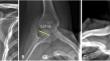
A prospective observational study was performed on 32 patients over two years at a tertiary orthopedic center. The loss of sclerotic glenoid rim (LSGL) on AP radiograph and the percentage relative glenoid bone loss on the Bernageau radiograph were assessed. The percentage glenoid bone loss and anterior straight line (ASL) were calculated using a best fit en face circle method using CT and MRI. Percentage glenoid bone loss was also calculated during arthroscopy in multiples of 5%.
Results
In our study, 90.6% (29) patients were males, while only 9.4% (3) were females. This can be attributed to the involvement of the males in outdoor activities and sports. Also, the maximum number of patients were found to belong to 21–30 years of age, with the mean age being 28.66 years. Of the 32 patients, loss of sclerotic glenoid line (LSGL) on AP radiographs correlated with glenoid bone loss on cross-sectional imaging in 27 patients. Three patients had equivocal LSGL and 2 patients with glenoid bone loss on CT did not demonstrate LSGL. The difference between the two modalities was not statistically significant (p value = 0.002). The glenoid bone loss on Bernageau view correlated with glenoid bone loss on cross sectional imaging in all but one patient. The bone loss as evaluated by radiograph Bernageau view was found to have strong correlation (correlation coefficient r = 0.948, p value < 0.0001).
Conclusion
AP and Bernageau radiographic views for anterior shoulder dislocations demonstrate good correlation with glenoid bone loss on cross-sectional imaging. They may also be used as an adjunct to predict overall bone loss on CT and at arthroscopy.
期刊介绍:
Skeletal Radiology provides a forum for the dissemination of current knowledge and information dealing with disorders of the musculoskeletal system including the spine. While emphasizing the radiological aspects of the many varied skeletal abnormalities, the journal also adopts an interdisciplinary approach, reflecting the membership of the International Skeletal Society. Thus, the anatomical, pathological, physiological, clinical, metabolic and epidemiological aspects of the many entities affecting the skeleton receive appropriate consideration.
This is the Journal of the International Skeletal Society and the Official Journal of the Society of Skeletal Radiology and the Australasian Musculoskelelal Imaging Group.


 分享
分享
 求助内容:
求助内容: 应助结果提醒方式:
 扫码关注我们
