Amine El Kandoussi, Yin P. Hung, Eric L. Tung, Fabian Bauer, Joao R. T. Vicentini, Santiago Lozano-Calderon, Connie Y. Chang
{"title":"骨外肌软骨肉瘤的临床、成像和病理特征","authors":"Amine El Kandoussi, Yin P. Hung, Eric L. Tung, Fabian Bauer, Joao R. T. Vicentini, Santiago Lozano-Calderon, Connie Y. Chang","doi":"10.1007/s00256-024-04800-6","DOIUrl":null,"url":null,"abstract":"<h3 data-test=\"abstract-sub-heading\">Objective</h3><p>To evaluate clinical and radiological features of extraskeletal myxoid chondrosarcomas (EMC).</p><h3 data-test=\"abstract-sub-heading\">Material and Methods</h3><p>Our pathology database was queried for cases of EMCs. Tumor location, size, imaging appearance, presence of metastases, disease recurrence, and clinical outcome were documented. Imaging studies were evaluated in consensus by a musculoskeletal radiologist and an orthopedic oncologist.</p><h3 data-test=\"abstract-sub-heading\">Results</h3><p>Thirty subjects met the inclusion criteria (mean age 52.7 ± 16.2 years; 19 male, 11 female), 17 (56.7%) of which had pre-operative imaging. Tumors occurred most often in the lower extremities (20/30; 66.7%). All cases presented as a soft-tissue mass without mineralization on XR or CT. On MRI, tumors were typically hyperintense on T2-weighted sequences (14/14; 100%) and had a chondroid matrix appearance (12/14; 85.7%). Tumor invasion was observed in 11 out of 16 (68.9%) patients and necrosis in 2 out of 11 subjects (18.2%). All subjects had their tumors examined by pathology, and 20 (66.7%) subjects also had descriptive information in addition to the diagnosis (tumor invasion, mitotic rate, and necrosis) noted in the pathology reports. The mean duration of follow-up was 9.4 ± 7.5 (1.0 – 29.6) years. At the last follow-up, 14 out of 28 (50%) subjects were disease-free, 6 out of 28 had persistent metastatic disease and 8 out of 28 had died.</p><h3 data-test=\"abstract-sub-heading\">Conclusions</h3><p>EMC is a rare sarcoma that commonly presents as lower extremity soft tissue mass with chondroid appearance on MRI. Unlike conventional chondrosarcomas, EMC do not demonstrate mineralization on XR or CT.</p>","PeriodicalId":21783,"journal":{"name":"Skeletal Radiology","volume":null,"pages":null},"PeriodicalIF":1.9000,"publicationDate":"2024-09-10","publicationTypes":"Journal Article","fieldsOfStudy":null,"isOpenAccess":false,"openAccessPdf":"","citationCount":"0","resultStr":"{\"title\":\"Clinical, imaging and pathological features of extraskeletal myxoid chondrosarcoma\",\"authors\":\"Amine El Kandoussi, Yin P. Hung, Eric L. Tung, Fabian Bauer, Joao R. T. Vicentini, Santiago Lozano-Calderon, Connie Y. Chang\",\"doi\":\"10.1007/s00256-024-04800-6\",\"DOIUrl\":null,\"url\":null,\"abstract\":\"<h3 data-test=\\\"abstract-sub-heading\\\">Objective</h3><p>To evaluate clinical and radiological features of extraskeletal myxoid chondrosarcomas (EMC).</p><h3 data-test=\\\"abstract-sub-heading\\\">Material and Methods</h3><p>Our pathology database was queried for cases of EMCs. Tumor location, size, imaging appearance, presence of metastases, disease recurrence, and clinical outcome were documented. Imaging studies were evaluated in consensus by a musculoskeletal radiologist and an orthopedic oncologist.</p><h3 data-test=\\\"abstract-sub-heading\\\">Results</h3><p>Thirty subjects met the inclusion criteria (mean age 52.7 ± 16.2 years; 19 male, 11 female), 17 (56.7%) of which had pre-operative imaging. Tumors occurred most often in the lower extremities (20/30; 66.7%). All cases presented as a soft-tissue mass without mineralization on XR or CT. On MRI, tumors were typically hyperintense on T2-weighted sequences (14/14; 100%) and had a chondroid matrix appearance (12/14; 85.7%). Tumor invasion was observed in 11 out of 16 (68.9%) patients and necrosis in 2 out of 11 subjects (18.2%). All subjects had their tumors examined by pathology, and 20 (66.7%) subjects also had descriptive information in addition to the diagnosis (tumor invasion, mitotic rate, and necrosis) noted in the pathology reports. The mean duration of follow-up was 9.4 ± 7.5 (1.0 – 29.6) years. At the last follow-up, 14 out of 28 (50%) subjects were disease-free, 6 out of 28 had persistent metastatic disease and 8 out of 28 had died.</p><h3 data-test=\\\"abstract-sub-heading\\\">Conclusions</h3><p>EMC is a rare sarcoma that commonly presents as lower extremity soft tissue mass with chondroid appearance on MRI. Unlike conventional chondrosarcomas, EMC do not demonstrate mineralization on XR or CT.</p>\",\"PeriodicalId\":21783,\"journal\":{\"name\":\"Skeletal Radiology\",\"volume\":null,\"pages\":null},\"PeriodicalIF\":1.9000,\"publicationDate\":\"2024-09-10\",\"publicationTypes\":\"Journal Article\",\"fieldsOfStudy\":null,\"isOpenAccess\":false,\"openAccessPdf\":\"\",\"citationCount\":\"0\",\"resultStr\":null,\"platform\":\"Semanticscholar\",\"paperid\":null,\"PeriodicalName\":\"Skeletal Radiology\",\"FirstCategoryId\":\"3\",\"ListUrlMain\":\"https://doi.org/10.1007/s00256-024-04800-6\",\"RegionNum\":3,\"RegionCategory\":\"医学\",\"ArticlePicture\":[],\"TitleCN\":null,\"AbstractTextCN\":null,\"PMCID\":null,\"EPubDate\":\"\",\"PubModel\":\"\",\"JCR\":\"Q2\",\"JCRName\":\"ORTHOPEDICS\",\"Score\":null,\"Total\":0}","platform":"Semanticscholar","paperid":null,"PeriodicalName":"Skeletal Radiology","FirstCategoryId":"3","ListUrlMain":"https://doi.org/10.1007/s00256-024-04800-6","RegionNum":3,"RegionCategory":"医学","ArticlePicture":[],"TitleCN":null,"AbstractTextCN":null,"PMCID":null,"EPubDate":"","PubModel":"","JCR":"Q2","JCRName":"ORTHOPEDICS","Score":null,"Total":0}
Clinical, imaging and pathological features of extraskeletal myxoid chondrosarcoma
Objective
To evaluate clinical and radiological features of extraskeletal myxoid chondrosarcomas (EMC).
Material and Methods
Our pathology database was queried for cases of EMCs. Tumor location, size, imaging appearance, presence of metastases, disease recurrence, and clinical outcome were documented. Imaging studies were evaluated in consensus by a musculoskeletal radiologist and an orthopedic oncologist.
Results
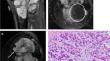
Thirty subjects met the inclusion criteria (mean age 52.7 ± 16.2 years; 19 male, 11 female), 17 (56.7%) of which had pre-operative imaging. Tumors occurred most often in the lower extremities (20/30; 66.7%). All cases presented as a soft-tissue mass without mineralization on XR or CT. On MRI, tumors were typically hyperintense on T2-weighted sequences (14/14; 100%) and had a chondroid matrix appearance (12/14; 85.7%). Tumor invasion was observed in 11 out of 16 (68.9%) patients and necrosis in 2 out of 11 subjects (18.2%). All subjects had their tumors examined by pathology, and 20 (66.7%) subjects also had descriptive information in addition to the diagnosis (tumor invasion, mitotic rate, and necrosis) noted in the pathology reports. The mean duration of follow-up was 9.4 ± 7.5 (1.0 – 29.6) years. At the last follow-up, 14 out of 28 (50%) subjects were disease-free, 6 out of 28 had persistent metastatic disease and 8 out of 28 had died.
Conclusions
EMC is a rare sarcoma that commonly presents as lower extremity soft tissue mass with chondroid appearance on MRI. Unlike conventional chondrosarcomas, EMC do not demonstrate mineralization on XR or CT.
期刊介绍:
Skeletal Radiology provides a forum for the dissemination of current knowledge and information dealing with disorders of the musculoskeletal system including the spine. While emphasizing the radiological aspects of the many varied skeletal abnormalities, the journal also adopts an interdisciplinary approach, reflecting the membership of the International Skeletal Society. Thus, the anatomical, pathological, physiological, clinical, metabolic and epidemiological aspects of the many entities affecting the skeleton receive appropriate consideration.
This is the Journal of the International Skeletal Society and the Official Journal of the Society of Skeletal Radiology and the Australasian Musculoskelelal Imaging Group.


 分享
分享
 求助内容:
求助内容: 应助结果提醒方式:
 扫码关注我们
