Wei Xue, Juanqin Niu, Gang Chen, Yao He, Xuesong Du, Fang Jingqin
{"title":"颞骨和颅底骨巨细胞瘤:6 例报告","authors":"Wei Xue, Juanqin Niu, Gang Chen, Yao He, Xuesong Du, Fang Jingqin","doi":"10.1007/s00256-024-04784-3","DOIUrl":null,"url":null,"abstract":"<h3 data-test=\"abstract-sub-heading\">Objective</h3><p>Five cases of giant cell tumor of bone (GCTB) in the head and neck region were reported, with a main focus on the radiological findings to identify common characteristics for the diagnosis of GCTB in these sites.</p><h3 data-test=\"abstract-sub-heading\">Materials and methods</h3><p>Five consecutive patients diagnosed with GCTB were retrospectively selected. Radiological features on conventional and advanced MR sequences and CT were analyzed. HE staining and immunohistochemical examination were performed using antibodies against p63 and CD68.</p><h3 data-test=\"abstract-sub-heading\">Results</h3><p>The common clinical features were local mass (3/5), tinnitus (3/5) and headache (2/5). Radiologically, all the cases were well-circumscribed osteolytic lesion, majority of cases demonstrated an expansile growth pattern and “soap bubble” appearance on CT (4/5). On MRI, the tumors showed predominantly hypointensity both on T1WI and T2WI, and no evidence of restricted diffusion on DWI. Intratumoral hemorrhage (2/5), cystic alternation (2/5) and very low signal on T2WI in the periphery region of the tumor (4/5) was found. Fluid–fluid level was noted in one case, which was eventually verified to be GCTB with secondary aneurysmal bone cyst (ABC). With contrast agent, all the cases showed striking (3/5) or mild to intermediate (2/5) enhancement.</p><h3 data-test=\"abstract-sub-heading\">Conclusions</h3><p>Although the above described radiological findings are not specific for GCTB in head and neck region, a well-defined osteolytic lesion in the bones of head and neck region with “soap bubble” appearance on CT and hypointensity on T2WI with very low signal in the peripheral region of the tumor on MRI highly suggest GCTB for patient ages 20 to 40.</p>","PeriodicalId":21783,"journal":{"name":"Skeletal Radiology","volume":"31 1","pages":""},"PeriodicalIF":2.2000,"publicationDate":"2024-09-02","publicationTypes":"Journal Article","fieldsOfStudy":null,"isOpenAccess":false,"openAccessPdf":"","citationCount":"0","resultStr":"{\"title\":\"Giant cell tumor of bone of temporal bone and skull base: report of 6 cases\",\"authors\":\"Wei Xue, Juanqin Niu, Gang Chen, Yao He, Xuesong Du, Fang Jingqin\",\"doi\":\"10.1007/s00256-024-04784-3\",\"DOIUrl\":null,\"url\":null,\"abstract\":\"<h3 data-test=\\\"abstract-sub-heading\\\">Objective</h3><p>Five cases of giant cell tumor of bone (GCTB) in the head and neck region were reported, with a main focus on the radiological findings to identify common characteristics for the diagnosis of GCTB in these sites.</p><h3 data-test=\\\"abstract-sub-heading\\\">Materials and methods</h3><p>Five consecutive patients diagnosed with GCTB were retrospectively selected. Radiological features on conventional and advanced MR sequences and CT were analyzed. HE staining and immunohistochemical examination were performed using antibodies against p63 and CD68.</p><h3 data-test=\\\"abstract-sub-heading\\\">Results</h3><p>The common clinical features were local mass (3/5), tinnitus (3/5) and headache (2/5). Radiologically, all the cases were well-circumscribed osteolytic lesion, majority of cases demonstrated an expansile growth pattern and “soap bubble” appearance on CT (4/5). On MRI, the tumors showed predominantly hypointensity both on T1WI and T2WI, and no evidence of restricted diffusion on DWI. Intratumoral hemorrhage (2/5), cystic alternation (2/5) and very low signal on T2WI in the periphery region of the tumor (4/5) was found. Fluid–fluid level was noted in one case, which was eventually verified to be GCTB with secondary aneurysmal bone cyst (ABC). With contrast agent, all the cases showed striking (3/5) or mild to intermediate (2/5) enhancement.</p><h3 data-test=\\\"abstract-sub-heading\\\">Conclusions</h3><p>Although the above described radiological findings are not specific for GCTB in head and neck region, a well-defined osteolytic lesion in the bones of head and neck region with “soap bubble” appearance on CT and hypointensity on T2WI with very low signal in the peripheral region of the tumor on MRI highly suggest GCTB for patient ages 20 to 40.</p>\",\"PeriodicalId\":21783,\"journal\":{\"name\":\"Skeletal Radiology\",\"volume\":\"31 1\",\"pages\":\"\"},\"PeriodicalIF\":2.2000,\"publicationDate\":\"2024-09-02\",\"publicationTypes\":\"Journal Article\",\"fieldsOfStudy\":null,\"isOpenAccess\":false,\"openAccessPdf\":\"\",\"citationCount\":\"0\",\"resultStr\":null,\"platform\":\"Semanticscholar\",\"paperid\":null,\"PeriodicalName\":\"Skeletal Radiology\",\"FirstCategoryId\":\"3\",\"ListUrlMain\":\"https://doi.org/10.1007/s00256-024-04784-3\",\"RegionNum\":3,\"RegionCategory\":\"医学\",\"ArticlePicture\":[],\"TitleCN\":null,\"AbstractTextCN\":null,\"PMCID\":null,\"EPubDate\":\"\",\"PubModel\":\"\",\"JCR\":\"Q2\",\"JCRName\":\"ORTHOPEDICS\",\"Score\":null,\"Total\":0}","platform":"Semanticscholar","paperid":null,"PeriodicalName":"Skeletal Radiology","FirstCategoryId":"3","ListUrlMain":"https://doi.org/10.1007/s00256-024-04784-3","RegionNum":3,"RegionCategory":"医学","ArticlePicture":[],"TitleCN":null,"AbstractTextCN":null,"PMCID":null,"EPubDate":"","PubModel":"","JCR":"Q2","JCRName":"ORTHOPEDICS","Score":null,"Total":0}
Giant cell tumor of bone of temporal bone and skull base: report of 6 cases
Objective
Five cases of giant cell tumor of bone (GCTB) in the head and neck region were reported, with a main focus on the radiological findings to identify common characteristics for the diagnosis of GCTB in these sites.
Materials and methods
Five consecutive patients diagnosed with GCTB were retrospectively selected. Radiological features on conventional and advanced MR sequences and CT were analyzed. HE staining and immunohistochemical examination were performed using antibodies against p63 and CD68.
Results
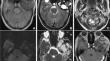
The common clinical features were local mass (3/5), tinnitus (3/5) and headache (2/5). Radiologically, all the cases were well-circumscribed osteolytic lesion, majority of cases demonstrated an expansile growth pattern and “soap bubble” appearance on CT (4/5). On MRI, the tumors showed predominantly hypointensity both on T1WI and T2WI, and no evidence of restricted diffusion on DWI. Intratumoral hemorrhage (2/5), cystic alternation (2/5) and very low signal on T2WI in the periphery region of the tumor (4/5) was found. Fluid–fluid level was noted in one case, which was eventually verified to be GCTB with secondary aneurysmal bone cyst (ABC). With contrast agent, all the cases showed striking (3/5) or mild to intermediate (2/5) enhancement.
Conclusions
Although the above described radiological findings are not specific for GCTB in head and neck region, a well-defined osteolytic lesion in the bones of head and neck region with “soap bubble” appearance on CT and hypointensity on T2WI with very low signal in the peripheral region of the tumor on MRI highly suggest GCTB for patient ages 20 to 40.
期刊介绍:
Skeletal Radiology provides a forum for the dissemination of current knowledge and information dealing with disorders of the musculoskeletal system including the spine. While emphasizing the radiological aspects of the many varied skeletal abnormalities, the journal also adopts an interdisciplinary approach, reflecting the membership of the International Skeletal Society. Thus, the anatomical, pathological, physiological, clinical, metabolic and epidemiological aspects of the many entities affecting the skeleton receive appropriate consideration.
This is the Journal of the International Skeletal Society and the Official Journal of the Society of Skeletal Radiology and the Australasian Musculoskelelal Imaging Group.


 分享
分享
 求助内容:
求助内容: 应助结果提醒方式:
 扫码关注我们
