{"title":"垂头综合征的放射学特征","authors":"Hiroshi Miyamoto","doi":"10.1007/s00586-024-08492-3","DOIUrl":null,"url":null,"abstract":"<h3 data-test=\"abstract-sub-heading\">Background</h3><p>This study aimed to elucidate the specificity of the radiological features of Dropped head syndrome (DHS) from both reginal and global aspects.</p><h3 data-test=\"abstract-sub-heading\">Methods</h3><p>We enrolled 53 patients with DHS (8 men, 45 women; mean age 73.5 years), and captured their lateral spinopelvic radiographs in standing position. We also selected 21 age- and sex-matched controls with cervical spondylosis. Radiological parameters were measured and compared between two groups. Compensatory and decompensatory sites were also listed for each patient.</p><h3 data-test=\"abstract-sub-heading\">Results</h3><p>Radiological factors such as sagittal vertical axis (SVA), clivo-axial angle (CAA), C2–7 angle, C2–7 SVA, anterior slippage of the vertebra, alignment. C1, C2, C3, C4, C5, C6 slopes, and T1 slope-C2–7 angle showed statistically significant differences between the groups. Multivariate logistic regression showed that SVA, C2–7 SVA, T1-slope-C2–7 angle, and C1 slope were the most important factors specific to DHS. Sole cervical spine and involvement of both cervical and thoracic spine accounted for 22% and 29% of the decompensatory sites in DHS respectively. Notably, 24% of the patients did not show decompensation of the cervical spine. While, 93% exhibited compensation at the craniovertebral junction. The thoracic spine contributed 70% to DHS compensation.</p><h3 data-test=\"abstract-sub-heading\">Conclusions</h3><p>This study indicated the radiological features of DHS from both regional and global aspects. Compensatory and decompensatory DHS mechanisms varied among individuals. Compensation was likely to be developed at the neighboring sites, with the craniovertebral and thoracic junctions as the proximal and distal parts for DHS, respectively.</p>","PeriodicalId":12323,"journal":{"name":"European Spine Journal","volume":"2 1","pages":""},"PeriodicalIF":2.7000,"publicationDate":"2024-09-12","publicationTypes":"Journal Article","fieldsOfStudy":null,"isOpenAccess":false,"openAccessPdf":"","citationCount":"0","resultStr":"{\"title\":\"Radiological features of dropped head syndrome\",\"authors\":\"Hiroshi Miyamoto\",\"doi\":\"10.1007/s00586-024-08492-3\",\"DOIUrl\":null,\"url\":null,\"abstract\":\"<h3 data-test=\\\"abstract-sub-heading\\\">Background</h3><p>This study aimed to elucidate the specificity of the radiological features of Dropped head syndrome (DHS) from both reginal and global aspects.</p><h3 data-test=\\\"abstract-sub-heading\\\">Methods</h3><p>We enrolled 53 patients with DHS (8 men, 45 women; mean age 73.5 years), and captured their lateral spinopelvic radiographs in standing position. We also selected 21 age- and sex-matched controls with cervical spondylosis. Radiological parameters were measured and compared between two groups. Compensatory and decompensatory sites were also listed for each patient.</p><h3 data-test=\\\"abstract-sub-heading\\\">Results</h3><p>Radiological factors such as sagittal vertical axis (SVA), clivo-axial angle (CAA), C2–7 angle, C2–7 SVA, anterior slippage of the vertebra, alignment. C1, C2, C3, C4, C5, C6 slopes, and T1 slope-C2–7 angle showed statistically significant differences between the groups. Multivariate logistic regression showed that SVA, C2–7 SVA, T1-slope-C2–7 angle, and C1 slope were the most important factors specific to DHS. Sole cervical spine and involvement of both cervical and thoracic spine accounted for 22% and 29% of the decompensatory sites in DHS respectively. Notably, 24% of the patients did not show decompensation of the cervical spine. While, 93% exhibited compensation at the craniovertebral junction. The thoracic spine contributed 70% to DHS compensation.</p><h3 data-test=\\\"abstract-sub-heading\\\">Conclusions</h3><p>This study indicated the radiological features of DHS from both regional and global aspects. Compensatory and decompensatory DHS mechanisms varied among individuals. Compensation was likely to be developed at the neighboring sites, with the craniovertebral and thoracic junctions as the proximal and distal parts for DHS, respectively.</p>\",\"PeriodicalId\":12323,\"journal\":{\"name\":\"European Spine Journal\",\"volume\":\"2 1\",\"pages\":\"\"},\"PeriodicalIF\":2.7000,\"publicationDate\":\"2024-09-12\",\"publicationTypes\":\"Journal Article\",\"fieldsOfStudy\":null,\"isOpenAccess\":false,\"openAccessPdf\":\"\",\"citationCount\":\"0\",\"resultStr\":null,\"platform\":\"Semanticscholar\",\"paperid\":null,\"PeriodicalName\":\"European Spine Journal\",\"FirstCategoryId\":\"3\",\"ListUrlMain\":\"https://doi.org/10.1007/s00586-024-08492-3\",\"RegionNum\":3,\"RegionCategory\":\"医学\",\"ArticlePicture\":[],\"TitleCN\":null,\"AbstractTextCN\":null,\"PMCID\":null,\"EPubDate\":\"\",\"PubModel\":\"\",\"JCR\":\"Q2\",\"JCRName\":\"CLINICAL NEUROLOGY\",\"Score\":null,\"Total\":0}","platform":"Semanticscholar","paperid":null,"PeriodicalName":"European Spine Journal","FirstCategoryId":"3","ListUrlMain":"https://doi.org/10.1007/s00586-024-08492-3","RegionNum":3,"RegionCategory":"医学","ArticlePicture":[],"TitleCN":null,"AbstractTextCN":null,"PMCID":null,"EPubDate":"","PubModel":"","JCR":"Q2","JCRName":"CLINICAL NEUROLOGY","Score":null,"Total":0}
This study aimed to elucidate the specificity of the radiological features of Dropped head syndrome (DHS) from both reginal and global aspects.
Methods
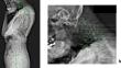
We enrolled 53 patients with DHS (8 men, 45 women; mean age 73.5 years), and captured their lateral spinopelvic radiographs in standing position. We also selected 21 age- and sex-matched controls with cervical spondylosis. Radiological parameters were measured and compared between two groups. Compensatory and decompensatory sites were also listed for each patient.
Results
Radiological factors such as sagittal vertical axis (SVA), clivo-axial angle (CAA), C2–7 angle, C2–7 SVA, anterior slippage of the vertebra, alignment. C1, C2, C3, C4, C5, C6 slopes, and T1 slope-C2–7 angle showed statistically significant differences between the groups. Multivariate logistic regression showed that SVA, C2–7 SVA, T1-slope-C2–7 angle, and C1 slope were the most important factors specific to DHS. Sole cervical spine and involvement of both cervical and thoracic spine accounted for 22% and 29% of the decompensatory sites in DHS respectively. Notably, 24% of the patients did not show decompensation of the cervical spine. While, 93% exhibited compensation at the craniovertebral junction. The thoracic spine contributed 70% to DHS compensation.
Conclusions
This study indicated the radiological features of DHS from both regional and global aspects. Compensatory and decompensatory DHS mechanisms varied among individuals. Compensation was likely to be developed at the neighboring sites, with the craniovertebral and thoracic junctions as the proximal and distal parts for DHS, respectively.
期刊介绍:
"European Spine Journal" is a publication founded in response to the increasing trend toward specialization in spinal surgery and spinal pathology in general. The Journal is devoted to all spine related disciplines, including functional and surgical anatomy of the spine, biomechanics and pathophysiology, diagnostic procedures, and neurology, surgery and outcomes. The aim of "European Spine Journal" is to support the further development of highly innovative spine treatments including but not restricted to surgery and to provide an integrated and balanced view of diagnostic, research and treatment procedures as well as outcomes that will enhance effective collaboration among specialists worldwide. The “European Spine Journal” also participates in education by means of videos, interactive meetings and the endorsement of educative efforts.
Official publication of EUROSPINE, The Spine Society of Europe


 分享
分享
 求助内容:
求助内容: 应助结果提醒方式:
 扫码关注我们
