Anna Timakova , Vladislav Ananev , Alexey Fayzullin , Egor Zemnuhov , Egor Rumyantsev , Andrey Zharov , Nicolay Zharkov , Varvara Zotova , Elena Shchelokova , Tatiana Demura , Peter Timashev , Vladimir Makarov
{"title":"LVI-PathNet:用于检测肺腺癌全切片图像中淋巴管侵犯的分割-分类管道","authors":"Anna Timakova , Vladislav Ananev , Alexey Fayzullin , Egor Zemnuhov , Egor Rumyantsev , Andrey Zharov , Nicolay Zharkov , Varvara Zotova , Elena Shchelokova , Tatiana Demura , Peter Timashev , Vladimir Makarov","doi":"10.1016/j.jpi.2024.100395","DOIUrl":null,"url":null,"abstract":"<div><p>Lymphovascular invasion (LVI) in lung cancer is a significant prognostic factor that influences treatment and outcomes, yet its reliable detection is challenging due to interobserver variability. This study aims to develop a deep learning model for LVI detection using whole slide images (WSIs) and evaluate its effectiveness within a pathologist's information system. Experienced pathologists annotated blood vessels and invading tumor cells in 162 WSIs of non-mucinous lung adenocarcinoma sourced from two external and one internal datasets. Two models were trained to segment vessels and identify images with LVI features. DeepLabV3+ model achieved an Intersection-over-Union of 0.8840 and an area under the receiver operating characteristic curve (AUC-ROC) of 0.9869 in vessel segmentation. For LVI classification, the ensemble model achieved a F1-score of 0.9683 and an AUC-ROC of 0.9987. The model demonstrated robustness and was unaffected by variations in staining and image quality. The pilot study showed that pathologists' evaluation time for LVI detecting decreased by an average of 16.95%, and by 21.5% in “hard cases”. The model facilitated consistent diagnostic assessments, suggesting potential for broader applications in detecting pathological changes in blood vessels and other lung pathologies.</p></div>","PeriodicalId":37769,"journal":{"name":"Journal of Pathology Informatics","volume":"15 ","pages":"Article 100395"},"PeriodicalIF":0.0000,"publicationDate":"2024-12-01","publicationTypes":"Journal Article","fieldsOfStudy":null,"isOpenAccess":false,"openAccessPdf":"https://www.sciencedirect.com/science/article/pii/S2153353924000348/pdfft?md5=9a1e9217891b1539c144069b2cb2703f&pid=1-s2.0-S2153353924000348-main.pdf","citationCount":"0","resultStr":"{\"title\":\"LVI-PathNet: Segmentation-classification pipeline for detection of lymphovascular invasion in whole slide images of lung adenocarcinoma\",\"authors\":\"Anna Timakova , Vladislav Ananev , Alexey Fayzullin , Egor Zemnuhov , Egor Rumyantsev , Andrey Zharov , Nicolay Zharkov , Varvara Zotova , Elena Shchelokova , Tatiana Demura , Peter Timashev , Vladimir Makarov\",\"doi\":\"10.1016/j.jpi.2024.100395\",\"DOIUrl\":null,\"url\":null,\"abstract\":\"<div><p>Lymphovascular invasion (LVI) in lung cancer is a significant prognostic factor that influences treatment and outcomes, yet its reliable detection is challenging due to interobserver variability. This study aims to develop a deep learning model for LVI detection using whole slide images (WSIs) and evaluate its effectiveness within a pathologist's information system. Experienced pathologists annotated blood vessels and invading tumor cells in 162 WSIs of non-mucinous lung adenocarcinoma sourced from two external and one internal datasets. Two models were trained to segment vessels and identify images with LVI features. DeepLabV3+ model achieved an Intersection-over-Union of 0.8840 and an area under the receiver operating characteristic curve (AUC-ROC) of 0.9869 in vessel segmentation. For LVI classification, the ensemble model achieved a F1-score of 0.9683 and an AUC-ROC of 0.9987. The model demonstrated robustness and was unaffected by variations in staining and image quality. The pilot study showed that pathologists' evaluation time for LVI detecting decreased by an average of 16.95%, and by 21.5% in “hard cases”. The model facilitated consistent diagnostic assessments, suggesting potential for broader applications in detecting pathological changes in blood vessels and other lung pathologies.</p></div>\",\"PeriodicalId\":37769,\"journal\":{\"name\":\"Journal of Pathology Informatics\",\"volume\":\"15 \",\"pages\":\"Article 100395\"},\"PeriodicalIF\":0.0000,\"publicationDate\":\"2024-12-01\",\"publicationTypes\":\"Journal Article\",\"fieldsOfStudy\":null,\"isOpenAccess\":false,\"openAccessPdf\":\"https://www.sciencedirect.com/science/article/pii/S2153353924000348/pdfft?md5=9a1e9217891b1539c144069b2cb2703f&pid=1-s2.0-S2153353924000348-main.pdf\",\"citationCount\":\"0\",\"resultStr\":null,\"platform\":\"Semanticscholar\",\"paperid\":null,\"PeriodicalName\":\"Journal of Pathology Informatics\",\"FirstCategoryId\":\"1085\",\"ListUrlMain\":\"https://www.sciencedirect.com/science/article/pii/S2153353924000348\",\"RegionNum\":0,\"RegionCategory\":null,\"ArticlePicture\":[],\"TitleCN\":null,\"AbstractTextCN\":null,\"PMCID\":null,\"EPubDate\":\"2024/8/30 0:00:00\",\"PubModel\":\"Epub\",\"JCR\":\"Q2\",\"JCRName\":\"Medicine\",\"Score\":null,\"Total\":0}","platform":"Semanticscholar","paperid":null,"PeriodicalName":"Journal of Pathology Informatics","FirstCategoryId":"1085","ListUrlMain":"https://www.sciencedirect.com/science/article/pii/S2153353924000348","RegionNum":0,"RegionCategory":null,"ArticlePicture":[],"TitleCN":null,"AbstractTextCN":null,"PMCID":null,"EPubDate":"2024/8/30 0:00:00","PubModel":"Epub","JCR":"Q2","JCRName":"Medicine","Score":null,"Total":0}
LVI-PathNet: Segmentation-classification pipeline for detection of lymphovascular invasion in whole slide images of lung adenocarcinoma
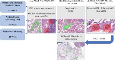
Lymphovascular invasion (LVI) in lung cancer is a significant prognostic factor that influences treatment and outcomes, yet its reliable detection is challenging due to interobserver variability. This study aims to develop a deep learning model for LVI detection using whole slide images (WSIs) and evaluate its effectiveness within a pathologist's information system. Experienced pathologists annotated blood vessels and invading tumor cells in 162 WSIs of non-mucinous lung adenocarcinoma sourced from two external and one internal datasets. Two models were trained to segment vessels and identify images with LVI features. DeepLabV3+ model achieved an Intersection-over-Union of 0.8840 and an area under the receiver operating characteristic curve (AUC-ROC) of 0.9869 in vessel segmentation. For LVI classification, the ensemble model achieved a F1-score of 0.9683 and an AUC-ROC of 0.9987. The model demonstrated robustness and was unaffected by variations in staining and image quality. The pilot study showed that pathologists' evaluation time for LVI detecting decreased by an average of 16.95%, and by 21.5% in “hard cases”. The model facilitated consistent diagnostic assessments, suggesting potential for broader applications in detecting pathological changes in blood vessels and other lung pathologies.
期刊介绍:
The Journal of Pathology Informatics (JPI) is an open access peer-reviewed journal dedicated to the advancement of pathology informatics. This is the official journal of the Association for Pathology Informatics (API). The journal aims to publish broadly about pathology informatics and freely disseminate all articles worldwide. This journal is of interest to pathologists, informaticians, academics, researchers, health IT specialists, information officers, IT staff, vendors, and anyone with an interest in informatics. We encourage submissions from anyone with an interest in the field of pathology informatics. We publish all types of papers related to pathology informatics including original research articles, technical notes, reviews, viewpoints, commentaries, editorials, symposia, meeting abstracts, book reviews, and correspondence to the editors. All submissions are subject to rigorous peer review by the well-regarded editorial board and by expert referees in appropriate specialties.


 分享
分享
 求助内容:
求助内容: 应助结果提醒方式:
 扫码关注我们
