Ingrid Framås Syversen, Daniel Reznik, Menno P. Witter, Asgeir Kobro-Flatmoen, Tobias Navarro Schröder, Christian F. Doeller
{"title":"优化后内侧与前外侧内侧皮层划分的 DTI-fMRI 联合方法。","authors":"Ingrid Framås Syversen, Daniel Reznik, Menno P. Witter, Asgeir Kobro-Flatmoen, Tobias Navarro Schröder, Christian F. Doeller","doi":"10.1002/hipo.23639","DOIUrl":null,"url":null,"abstract":"<p>In the entorhinal cortex (EC), attempts have been made to identify the human homologue regions of the medial (MEC) and lateral (LEC) subregions using either functional magnetic resonance imaging (fMRI) or diffusion tensor imaging (DTI). However, there are still discrepancies between entorhinal subdivisions depending on the choice of connectivity seed regions and the imaging modality used. While DTI can be used to follow the white matter tracts of the brain, fMRI can identify functionally connected brain regions. In this study, we used both DTI and resting-state fMRI in 103 healthy adults to investigate both structural and functional connectivity between the EC and associated cortical brain regions. Differential connectivity with these regions was then used to predict the locations of the human homologues of MEC and LEC. Our results from combining DTI and fMRI support a subdivision into posteromedial (pmEC) and anterolateral (alEC) EC and reveal a confined border between the pmEC and alEC. Furthermore, the EC subregions obtained by either imaging modality showed similar distinct whole-brain connectivity profiles. Optimizing the delineation of the human homologues of MEC and LEC with a combined, cross-validated DTI-fMRI approach allows to define a likely border between the two subdivisions and has implications for both cognitive and translational neuroscience research.</p>","PeriodicalId":13171,"journal":{"name":"Hippocampus","volume":"34 11","pages":"659-672"},"PeriodicalIF":2.7000,"publicationDate":"2024-09-21","publicationTypes":"Journal Article","fieldsOfStudy":null,"isOpenAccess":false,"openAccessPdf":"https://onlinelibrary.wiley.com/doi/epdf/10.1002/hipo.23639","citationCount":"0","resultStr":"{\"title\":\"A combined DTI-fMRI approach for optimizing the delineation of posteromedial versus anterolateral entorhinal cortex\",\"authors\":\"Ingrid Framås Syversen, Daniel Reznik, Menno P. Witter, Asgeir Kobro-Flatmoen, Tobias Navarro Schröder, Christian F. Doeller\",\"doi\":\"10.1002/hipo.23639\",\"DOIUrl\":null,\"url\":null,\"abstract\":\"<p>In the entorhinal cortex (EC), attempts have been made to identify the human homologue regions of the medial (MEC) and lateral (LEC) subregions using either functional magnetic resonance imaging (fMRI) or diffusion tensor imaging (DTI). However, there are still discrepancies between entorhinal subdivisions depending on the choice of connectivity seed regions and the imaging modality used. While DTI can be used to follow the white matter tracts of the brain, fMRI can identify functionally connected brain regions. In this study, we used both DTI and resting-state fMRI in 103 healthy adults to investigate both structural and functional connectivity between the EC and associated cortical brain regions. Differential connectivity with these regions was then used to predict the locations of the human homologues of MEC and LEC. Our results from combining DTI and fMRI support a subdivision into posteromedial (pmEC) and anterolateral (alEC) EC and reveal a confined border between the pmEC and alEC. Furthermore, the EC subregions obtained by either imaging modality showed similar distinct whole-brain connectivity profiles. Optimizing the delineation of the human homologues of MEC and LEC with a combined, cross-validated DTI-fMRI approach allows to define a likely border between the two subdivisions and has implications for both cognitive and translational neuroscience research.</p>\",\"PeriodicalId\":13171,\"journal\":{\"name\":\"Hippocampus\",\"volume\":\"34 11\",\"pages\":\"659-672\"},\"PeriodicalIF\":2.7000,\"publicationDate\":\"2024-09-21\",\"publicationTypes\":\"Journal Article\",\"fieldsOfStudy\":null,\"isOpenAccess\":false,\"openAccessPdf\":\"https://onlinelibrary.wiley.com/doi/epdf/10.1002/hipo.23639\",\"citationCount\":\"0\",\"resultStr\":null,\"platform\":\"Semanticscholar\",\"paperid\":null,\"PeriodicalName\":\"Hippocampus\",\"FirstCategoryId\":\"3\",\"ListUrlMain\":\"https://onlinelibrary.wiley.com/doi/10.1002/hipo.23639\",\"RegionNum\":3,\"RegionCategory\":\"医学\",\"ArticlePicture\":[],\"TitleCN\":null,\"AbstractTextCN\":null,\"PMCID\":null,\"EPubDate\":\"\",\"PubModel\":\"\",\"JCR\":\"Q3\",\"JCRName\":\"NEUROSCIENCES\",\"Score\":null,\"Total\":0}","platform":"Semanticscholar","paperid":null,"PeriodicalName":"Hippocampus","FirstCategoryId":"3","ListUrlMain":"https://onlinelibrary.wiley.com/doi/10.1002/hipo.23639","RegionNum":3,"RegionCategory":"医学","ArticlePicture":[],"TitleCN":null,"AbstractTextCN":null,"PMCID":null,"EPubDate":"","PubModel":"","JCR":"Q3","JCRName":"NEUROSCIENCES","Score":null,"Total":0}
A combined DTI-fMRI approach for optimizing the delineation of posteromedial versus anterolateral entorhinal cortex
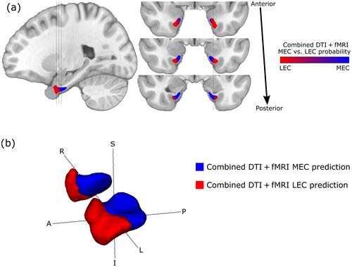
In the entorhinal cortex (EC), attempts have been made to identify the human homologue regions of the medial (MEC) and lateral (LEC) subregions using either functional magnetic resonance imaging (fMRI) or diffusion tensor imaging (DTI). However, there are still discrepancies between entorhinal subdivisions depending on the choice of connectivity seed regions and the imaging modality used. While DTI can be used to follow the white matter tracts of the brain, fMRI can identify functionally connected brain regions. In this study, we used both DTI and resting-state fMRI in 103 healthy adults to investigate both structural and functional connectivity between the EC and associated cortical brain regions. Differential connectivity with these regions was then used to predict the locations of the human homologues of MEC and LEC. Our results from combining DTI and fMRI support a subdivision into posteromedial (pmEC) and anterolateral (alEC) EC and reveal a confined border between the pmEC and alEC. Furthermore, the EC subregions obtained by either imaging modality showed similar distinct whole-brain connectivity profiles. Optimizing the delineation of the human homologues of MEC and LEC with a combined, cross-validated DTI-fMRI approach allows to define a likely border between the two subdivisions and has implications for both cognitive and translational neuroscience research.
期刊介绍:
Hippocampus provides a forum for the exchange of current information between investigators interested in the neurobiology of the hippocampal formation and related structures. While the relationships of submitted papers to the hippocampal formation will be evaluated liberally, the substance of appropriate papers should deal with the hippocampal formation per se or with the interaction between the hippocampal formation and other brain regions. The scope of Hippocampus is wide: single and multidisciplinary experimental studies from all fields of basic science, theoretical papers, papers dealing with hippocampal preparations as models for understanding the central nervous system, and clinical studies will be considered for publication. The Editor especially encourages the submission of papers that contribute to a functional understanding of the hippocampal formation.


 分享
分享
 求助内容:
求助内容: 应助结果提醒方式:
 扫码关注我们
