Rada Ellegård , Torsten Malm , Constance G. Weismann , Eva Fernlund , Swedish National Biobank for Congenital Heart Disease (SNAB-CHD) consortium, Anneli Nordén Björnlert , Hanna Klang Årstrand , Katarina Ellnebo-Svedlund , Cecilia Gunnarsson
{"title":"主动脉闭塞区转录组分析","authors":"Rada Ellegård , Torsten Malm , Constance G. Weismann , Eva Fernlund , Swedish National Biobank for Congenital Heart Disease (SNAB-CHD) consortium, Anneli Nordén Björnlert , Hanna Klang Årstrand , Katarina Ellnebo-Svedlund , Cecilia Gunnarsson","doi":"10.1016/j.jmccpl.2024.100094","DOIUrl":null,"url":null,"abstract":"<div><h3>Background</h3><div>Coarctation of the aorta (CoA) is a relatively common congenital heart defect. The underlying causes are not known, but a combination of genetic factors and abnormalities linked to embryonic development is suspected. There are only a few studies of the underlying molecular mechanisms in CoA. The aim of the current study was to expand our understanding of the pathogenesis of CoA by characterizing the transcriptome of the coarctation area.</div></div><div><h3>Methods</h3><div>Tissue samples from 21 pediatric patients operated for CoA were dissected into separate biopsies consisting of the localized coarctation itself, proximal/distal tissue and ductus. RNA was sequenced to evaluate gene expression in the different biopsies.</div></div><div><h3>Results</h3><div>We observed an activation of acute phase response in samples from the localized coarctation compared to samples from distal or proximal tissue. However, we observed even bigger differences for patient age and sex than compared to biopsy location. A cluster of genes located at 1q21, including the S100 gene family, displayed contrasting expression depending on patient sex, and appeared to affect the balance between inflammatory and interferon pathways. Biopsies from patients <3 months old were characterized by a significantly higher fibrotic activity compared to samples from older patients. The ductus tissue was characterized by an upregulation of factors associated with proliferation.</div></div><div><h3>Conclusions</h3><div>The ongoing processes in the coarctation area are influenced by the age and sex of the patient, and possibly by differences in etiology between different patients. The impact of patient attributes must be taken into consideration when performing future studies.</div></div>","PeriodicalId":73835,"journal":{"name":"Journal of molecular and cellular cardiology plus","volume":"10 ","pages":"Article 100094"},"PeriodicalIF":2.2000,"publicationDate":"2024-12-01","publicationTypes":"Journal Article","fieldsOfStudy":null,"isOpenAccess":false,"openAccessPdf":"https://www.sciencedirect.com/science/article/pii/S2772976124000345/pdfft?md5=c075465d71ff736e6bcc8d95cc7ac49d&pid=1-s2.0-S2772976124000345-main.pdf","citationCount":"0","resultStr":"{\"title\":\"Transcriptome analysis of the aortic coarctation area\",\"authors\":\"Rada Ellegård , Torsten Malm , Constance G. Weismann , Eva Fernlund , Swedish National Biobank for Congenital Heart Disease (SNAB-CHD) consortium, Anneli Nordén Björnlert , Hanna Klang Årstrand , Katarina Ellnebo-Svedlund , Cecilia Gunnarsson\",\"doi\":\"10.1016/j.jmccpl.2024.100094\",\"DOIUrl\":null,\"url\":null,\"abstract\":\"<div><h3>Background</h3><div>Coarctation of the aorta (CoA) is a relatively common congenital heart defect. The underlying causes are not known, but a combination of genetic factors and abnormalities linked to embryonic development is suspected. There are only a few studies of the underlying molecular mechanisms in CoA. The aim of the current study was to expand our understanding of the pathogenesis of CoA by characterizing the transcriptome of the coarctation area.</div></div><div><h3>Methods</h3><div>Tissue samples from 21 pediatric patients operated for CoA were dissected into separate biopsies consisting of the localized coarctation itself, proximal/distal tissue and ductus. RNA was sequenced to evaluate gene expression in the different biopsies.</div></div><div><h3>Results</h3><div>We observed an activation of acute phase response in samples from the localized coarctation compared to samples from distal or proximal tissue. However, we observed even bigger differences for patient age and sex than compared to biopsy location. A cluster of genes located at 1q21, including the S100 gene family, displayed contrasting expression depending on patient sex, and appeared to affect the balance between inflammatory and interferon pathways. Biopsies from patients <3 months old were characterized by a significantly higher fibrotic activity compared to samples from older patients. The ductus tissue was characterized by an upregulation of factors associated with proliferation.</div></div><div><h3>Conclusions</h3><div>The ongoing processes in the coarctation area are influenced by the age and sex of the patient, and possibly by differences in etiology between different patients. The impact of patient attributes must be taken into consideration when performing future studies.</div></div>\",\"PeriodicalId\":73835,\"journal\":{\"name\":\"Journal of molecular and cellular cardiology plus\",\"volume\":\"10 \",\"pages\":\"Article 100094\"},\"PeriodicalIF\":2.2000,\"publicationDate\":\"2024-12-01\",\"publicationTypes\":\"Journal Article\",\"fieldsOfStudy\":null,\"isOpenAccess\":false,\"openAccessPdf\":\"https://www.sciencedirect.com/science/article/pii/S2772976124000345/pdfft?md5=c075465d71ff736e6bcc8d95cc7ac49d&pid=1-s2.0-S2772976124000345-main.pdf\",\"citationCount\":\"0\",\"resultStr\":null,\"platform\":\"Semanticscholar\",\"paperid\":null,\"PeriodicalName\":\"Journal of molecular and cellular cardiology plus\",\"FirstCategoryId\":\"1085\",\"ListUrlMain\":\"https://www.sciencedirect.com/science/article/pii/S2772976124000345\",\"RegionNum\":0,\"RegionCategory\":null,\"ArticlePicture\":[],\"TitleCN\":null,\"AbstractTextCN\":null,\"PMCID\":null,\"EPubDate\":\"2024/9/17 0:00:00\",\"PubModel\":\"Epub\",\"JCR\":\"\",\"JCRName\":\"\",\"Score\":null,\"Total\":0}","platform":"Semanticscholar","paperid":null,"PeriodicalName":"Journal of molecular and cellular cardiology plus","FirstCategoryId":"1085","ListUrlMain":"https://www.sciencedirect.com/science/article/pii/S2772976124000345","RegionNum":0,"RegionCategory":null,"ArticlePicture":[],"TitleCN":null,"AbstractTextCN":null,"PMCID":null,"EPubDate":"2024/9/17 0:00:00","PubModel":"Epub","JCR":"","JCRName":"","Score":null,"Total":0}
引用次数: 0
摘要
背景主动脉瘤(CoA)是一种比较常见的先天性心脏缺陷。其根本原因尚不清楚,但怀疑与遗传因素和胚胎发育异常有关。目前只有少数几项关于 CoA 潜在分子机制的研究。本研究的目的是通过分析冠状动脉畸形区域转录组的特征,扩大我们对 CoA 发病机制的了解。方法:将 21 例因 CoA 而接受手术的儿科患者的组织样本分成不同的活检组织,包括局部冠状动脉畸形本身、近端/远端组织和导管。我们观察到,与来自远端或近端组织的样本相比,来自局部冠状动脉的样本中的急性期反应被激活。然而,与活检位置相比,我们发现患者年龄和性别的差异更大。位于 1q21 的一组基因(包括 S100 基因家族)的表达因患者性别而异,似乎影响了炎症和干扰素途径之间的平衡。与年龄较大的患者样本相比,3 个月大的患者活检样本的纤维化活性明显更高。导管组织的特点是与增殖相关的因子上调。在今后的研究中必须考虑到患者属性的影响。
Transcriptome analysis of the aortic coarctation area
Background
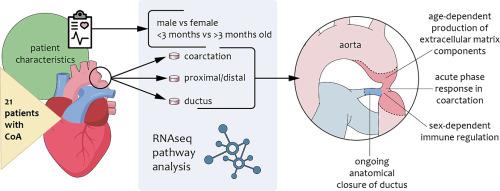
Coarctation of the aorta (CoA) is a relatively common congenital heart defect. The underlying causes are not known, but a combination of genetic factors and abnormalities linked to embryonic development is suspected. There are only a few studies of the underlying molecular mechanisms in CoA. The aim of the current study was to expand our understanding of the pathogenesis of CoA by characterizing the transcriptome of the coarctation area.
Methods
Tissue samples from 21 pediatric patients operated for CoA were dissected into separate biopsies consisting of the localized coarctation itself, proximal/distal tissue and ductus. RNA was sequenced to evaluate gene expression in the different biopsies.
Results
We observed an activation of acute phase response in samples from the localized coarctation compared to samples from distal or proximal tissue. However, we observed even bigger differences for patient age and sex than compared to biopsy location. A cluster of genes located at 1q21, including the S100 gene family, displayed contrasting expression depending on patient sex, and appeared to affect the balance between inflammatory and interferon pathways. Biopsies from patients <3 months old were characterized by a significantly higher fibrotic activity compared to samples from older patients. The ductus tissue was characterized by an upregulation of factors associated with proliferation.
Conclusions
The ongoing processes in the coarctation area are influenced by the age and sex of the patient, and possibly by differences in etiology between different patients. The impact of patient attributes must be taken into consideration when performing future studies.


 分享
分享
 求助内容:
求助内容: 应助结果提醒方式:
 扫码关注我们
