Amr Abdin, Alaa Din Abdin, Giuseppe Merone, Wissam Aljundi, Bernhard Haring, Yaser Abu Dail, Felix Mahfoud, Insa Emrich, Hussam Al Ghorani, Elsa Wilma Böhm, Berthold Seitz, Michael Böhm
{"title":"心眼综合征:急性失代偿性心力衰竭的视网膜微血管变化","authors":"Amr Abdin, Alaa Din Abdin, Giuseppe Merone, Wissam Aljundi, Bernhard Haring, Yaser Abu Dail, Felix Mahfoud, Insa Emrich, Hussam Al Ghorani, Elsa Wilma Böhm, Berthold Seitz, Michael Böhm","doi":"10.1002/ejhf.3474","DOIUrl":null,"url":null,"abstract":"<div>\n \n \n <section>\n \n <h3> Aims</h3>\n \n <p>To investigate the changes in retinal microvasculature by contemporary imaging techniques during episodes of acute decompensated heart failure (ADHF) and following recompensation compared to age-matched controls without known cardiac or retinal disease.</p>\n </section>\n \n <section>\n \n <h3> Methods and results</h3>\n \n <p>Adult patients hospitalized with a primary diagnosis of ADHF, regardless of left ventricular ejection fraction (LVEF) and treated with a minimum dose of 40 mg of intravenous furosemide or equivalent were included. Transthoracic echocardiography was conducted in all patients. Eye examinations were performed out within the initial 24 h after admission and after recompensation before discharge. All eyes underwent a general examination, including a best corrected visual acuity test, dilated fundoscopy, spectral-domain optical coherence tomography (OCT) as well as OCT angiography (OCT-A). In addition, 40 participants without documented cardiac or retinal diseases served as controls. Forty patients with ADHF (mean age 78.9 ± 8.8 years; 32% female) with a mean LVEF of 43 ± 12.8% were included. All patients were treated with intravenous diuretics for a median of 4.3 ± 2.8 days. There was a significant reduction in N-terminal pro-B-type natriuretic peptide from baseline up to discharge (10 396 [interquartile range 6410] vs. 6380 [interquartile range 3933] pg/ml, <i>p</i> ≤ 0.001) and inferior vena cava diameters (2.13 ± 0.4 vs. 1.63 ± 0.3 cm, <i>p</i> = 0.003). Compared to the control group, patients with ADHF showed on admission impaired visual acuity (0.15 ± 0.1 vs. 0.35 ± 0.1 logMAR, <i>p</i> < 0.001), reduced macular vessel density (18.0 ± 1.9 vs. 14.3 ± 3.6 mm/mm<sup>2</sup>, <i>p</i> < 0.001) and perfusion density (42.6 ± 3.2 vs. 35.2 ± 9.7%, <i>p</i> < 0.001). After recompensation, the mean overall vessel density and mean overall perfusion density were markedly increased at discharge (14.3 ± 3.6 vs. 19.7 ± 2.6 mm/mm<sup>2</sup>, <i>p</i> = 0.001, and 35.2 ± 9.7 vs. 39.2 ± 6.5%, <i>p</i> = 0.005, respectively). The mean diameter of the superior temporal retinal vein at admission was significantly larger compared to the control group (136 ± 19 vs. 124 ± 22 μm, <i>p</i> = 0.008) and decreased significantly to 122 ± 15 μm at discharge (<i>p</i> < 0.001).</p>\n </section>\n \n <section>\n \n <h3> Conclusion</h3>\n \n <p>This analysis revealed a remarkable reversible change in retinal microvasculature after ADHF. This could provide a valuable evidence for use of OCT-A in the assessment of overall microperfusion and haemodynamic status in patients with acute heart failure.</p>\n </section>\n </div>","PeriodicalId":164,"journal":{"name":"European Journal of Heart Failure","volume":"26 11","pages":"2421-2430"},"PeriodicalIF":10.3000,"publicationDate":"2024-09-24","publicationTypes":"Journal Article","fieldsOfStudy":null,"isOpenAccess":false,"openAccessPdf":"https://onlinelibrary.wiley.com/doi/epdf/10.1002/ejhf.3474","citationCount":"0","resultStr":"{\"title\":\"Cardio-ocular syndrome: Retinal microvascular changes in acutely decompensated heart failure\",\"authors\":\"Amr Abdin, Alaa Din Abdin, Giuseppe Merone, Wissam Aljundi, Bernhard Haring, Yaser Abu Dail, Felix Mahfoud, Insa Emrich, Hussam Al Ghorani, Elsa Wilma Böhm, Berthold Seitz, Michael Böhm\",\"doi\":\"10.1002/ejhf.3474\",\"DOIUrl\":null,\"url\":null,\"abstract\":\"<div>\\n \\n \\n <section>\\n \\n <h3> Aims</h3>\\n \\n <p>To investigate the changes in retinal microvasculature by contemporary imaging techniques during episodes of acute decompensated heart failure (ADHF) and following recompensation compared to age-matched controls without known cardiac or retinal disease.</p>\\n </section>\\n \\n <section>\\n \\n <h3> Methods and results</h3>\\n \\n <p>Adult patients hospitalized with a primary diagnosis of ADHF, regardless of left ventricular ejection fraction (LVEF) and treated with a minimum dose of 40 mg of intravenous furosemide or equivalent were included. Transthoracic echocardiography was conducted in all patients. Eye examinations were performed out within the initial 24 h after admission and after recompensation before discharge. All eyes underwent a general examination, including a best corrected visual acuity test, dilated fundoscopy, spectral-domain optical coherence tomography (OCT) as well as OCT angiography (OCT-A). In addition, 40 participants without documented cardiac or retinal diseases served as controls. Forty patients with ADHF (mean age 78.9 ± 8.8 years; 32% female) with a mean LVEF of 43 ± 12.8% were included. All patients were treated with intravenous diuretics for a median of 4.3 ± 2.8 days. There was a significant reduction in N-terminal pro-B-type natriuretic peptide from baseline up to discharge (10 396 [interquartile range 6410] vs. 6380 [interquartile range 3933] pg/ml, <i>p</i> ≤ 0.001) and inferior vena cava diameters (2.13 ± 0.4 vs. 1.63 ± 0.3 cm, <i>p</i> = 0.003). Compared to the control group, patients with ADHF showed on admission impaired visual acuity (0.15 ± 0.1 vs. 0.35 ± 0.1 logMAR, <i>p</i> < 0.001), reduced macular vessel density (18.0 ± 1.9 vs. 14.3 ± 3.6 mm/mm<sup>2</sup>, <i>p</i> < 0.001) and perfusion density (42.6 ± 3.2 vs. 35.2 ± 9.7%, <i>p</i> < 0.001). After recompensation, the mean overall vessel density and mean overall perfusion density were markedly increased at discharge (14.3 ± 3.6 vs. 19.7 ± 2.6 mm/mm<sup>2</sup>, <i>p</i> = 0.001, and 35.2 ± 9.7 vs. 39.2 ± 6.5%, <i>p</i> = 0.005, respectively). The mean diameter of the superior temporal retinal vein at admission was significantly larger compared to the control group (136 ± 19 vs. 124 ± 22 μm, <i>p</i> = 0.008) and decreased significantly to 122 ± 15 μm at discharge (<i>p</i> < 0.001).</p>\\n </section>\\n \\n <section>\\n \\n <h3> Conclusion</h3>\\n \\n <p>This analysis revealed a remarkable reversible change in retinal microvasculature after ADHF. This could provide a valuable evidence for use of OCT-A in the assessment of overall microperfusion and haemodynamic status in patients with acute heart failure.</p>\\n </section>\\n </div>\",\"PeriodicalId\":164,\"journal\":{\"name\":\"European Journal of Heart Failure\",\"volume\":\"26 11\",\"pages\":\"2421-2430\"},\"PeriodicalIF\":10.3000,\"publicationDate\":\"2024-09-24\",\"publicationTypes\":\"Journal Article\",\"fieldsOfStudy\":null,\"isOpenAccess\":false,\"openAccessPdf\":\"https://onlinelibrary.wiley.com/doi/epdf/10.1002/ejhf.3474\",\"citationCount\":\"0\",\"resultStr\":null,\"platform\":\"Semanticscholar\",\"paperid\":null,\"PeriodicalName\":\"European Journal of Heart Failure\",\"FirstCategoryId\":\"3\",\"ListUrlMain\":\"https://onlinelibrary.wiley.com/doi/10.1002/ejhf.3474\",\"RegionNum\":1,\"RegionCategory\":\"医学\",\"ArticlePicture\":[],\"TitleCN\":null,\"AbstractTextCN\":null,\"PMCID\":null,\"EPubDate\":\"\",\"PubModel\":\"\",\"JCR\":\"Q1\",\"JCRName\":\"CARDIAC & CARDIOVASCULAR SYSTEMS\",\"Score\":null,\"Total\":0}","platform":"Semanticscholar","paperid":null,"PeriodicalName":"European Journal of Heart Failure","FirstCategoryId":"3","ListUrlMain":"https://onlinelibrary.wiley.com/doi/10.1002/ejhf.3474","RegionNum":1,"RegionCategory":"医学","ArticlePicture":[],"TitleCN":null,"AbstractTextCN":null,"PMCID":null,"EPubDate":"","PubModel":"","JCR":"Q1","JCRName":"CARDIAC & CARDIOVASCULAR SYSTEMS","Score":null,"Total":0}
Cardio-ocular syndrome: Retinal microvascular changes in acutely decompensated heart failure
Aims
To investigate the changes in retinal microvasculature by contemporary imaging techniques during episodes of acute decompensated heart failure (ADHF) and following recompensation compared to age-matched controls without known cardiac or retinal disease.
Methods and results
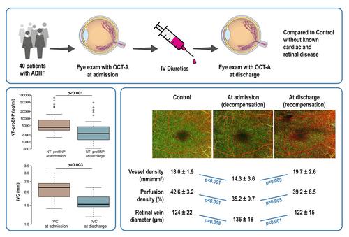
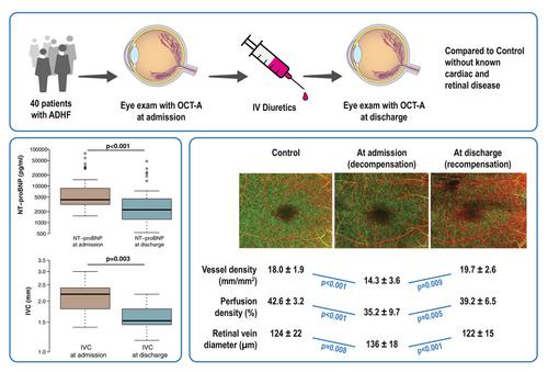
Adult patients hospitalized with a primary diagnosis of ADHF, regardless of left ventricular ejection fraction (LVEF) and treated with a minimum dose of 40 mg of intravenous furosemide or equivalent were included. Transthoracic echocardiography was conducted in all patients. Eye examinations were performed out within the initial 24 h after admission and after recompensation before discharge. All eyes underwent a general examination, including a best corrected visual acuity test, dilated fundoscopy, spectral-domain optical coherence tomography (OCT) as well as OCT angiography (OCT-A). In addition, 40 participants without documented cardiac or retinal diseases served as controls. Forty patients with ADHF (mean age 78.9 ± 8.8 years; 32% female) with a mean LVEF of 43 ± 12.8% were included. All patients were treated with intravenous diuretics for a median of 4.3 ± 2.8 days. There was a significant reduction in N-terminal pro-B-type natriuretic peptide from baseline up to discharge (10 396 [interquartile range 6410] vs. 6380 [interquartile range 3933] pg/ml, p ≤ 0.001) and inferior vena cava diameters (2.13 ± 0.4 vs. 1.63 ± 0.3 cm, p = 0.003). Compared to the control group, patients with ADHF showed on admission impaired visual acuity (0.15 ± 0.1 vs. 0.35 ± 0.1 logMAR, p < 0.001), reduced macular vessel density (18.0 ± 1.9 vs. 14.3 ± 3.6 mm/mm2, p < 0.001) and perfusion density (42.6 ± 3.2 vs. 35.2 ± 9.7%, p < 0.001). After recompensation, the mean overall vessel density and mean overall perfusion density were markedly increased at discharge (14.3 ± 3.6 vs. 19.7 ± 2.6 mm/mm2, p = 0.001, and 35.2 ± 9.7 vs. 39.2 ± 6.5%, p = 0.005, respectively). The mean diameter of the superior temporal retinal vein at admission was significantly larger compared to the control group (136 ± 19 vs. 124 ± 22 μm, p = 0.008) and decreased significantly to 122 ± 15 μm at discharge (p < 0.001).
Conclusion
This analysis revealed a remarkable reversible change in retinal microvasculature after ADHF. This could provide a valuable evidence for use of OCT-A in the assessment of overall microperfusion and haemodynamic status in patients with acute heart failure.
期刊介绍:
European Journal of Heart Failure is an international journal dedicated to advancing knowledge in the field of heart failure management. The journal publishes reviews and editorials aimed at improving understanding, prevention, investigation, and treatment of heart failure. It covers various disciplines such as molecular and cellular biology, pathology, physiology, electrophysiology, pharmacology, clinical sciences, social sciences, and population sciences. The journal welcomes submissions of manuscripts on basic, clinical, and population sciences, as well as original contributions on nursing, care of the elderly, primary care, health economics, and other related specialist fields. It is published monthly and has a readership that includes cardiologists, emergency room physicians, intensivists, internists, general physicians, cardiac nurses, diabetologists, epidemiologists, basic scientists focusing on cardiovascular research, and those working in rehabilitation. The journal is abstracted and indexed in various databases such as Academic Search, Embase, MEDLINE/PubMed, and Science Citation Index.




 京公网安备 11010802042870号
京ICP备2023020795号-1
京公网安备 11010802042870号
京ICP备2023020795号-1

 分享
分享
 求助内容:
求助内容: 应助结果提醒方式:
 扫码关注我们
