Daniil Nozdriukhin, Marco Cattaneo, Norman Klingler, Shuxin Lyu, Weiye Li, Francisco Montero de Espinosa, Jerome Bonvin, Outi Supponen, Daniel Razansky, Xosé Luís Deán-Ben
{"title":"纳米多孔亚微米金颗粒实现了基于纳米颗粒的定位光声断层扫描(nanoLOT)","authors":"Daniil Nozdriukhin, Marco Cattaneo, Norman Klingler, Shuxin Lyu, Weiye Li, Francisco Montero de Espinosa, Jerome Bonvin, Outi Supponen, Daniel Razansky, Xosé Luís Deán-Ben","doi":"10.1002/smll.202404904","DOIUrl":null,"url":null,"abstract":"<p>Localization optoacoustic tomography (LOT) has recently emerged as a transformative super-resolution technique breaking through the acoustic diffraction limit in deep-tissue optoacoustic (OA) imaging via individual localization and tracking of particles in the bloodstream. However, strong light absorption in red blood cells has previously restricted per-particle OA detection to relatively large microparticles, ≈5 µm in diameter. Herein, it is demonstrated that submicron-sized porous gold nanoparticles, ≈600 nm in diameter, can be individually detected for noninvasive super-resolution imaging with LOT. Ultra-high-speed bright-field microscopy revealed that these nanoparticles generate microscopic plasmonic vapor bubbles, significantly enhancing opto-acoustic energy conversion through a nano-to-micro size transformation. Comprehensive in vitro and in vivo tests further demonstrated the biocompatibility and biosafety of the particles. By reducing the detectable particle size by an order of magnitude, nanoLOT enables microangiographic imaging with a significantly reduced risk of embolisms from particle aggregation and opens new avenues to visualize how nanoparticles reach vascular and potentially extravascular targets. The performance of nanoLOT for non-invasive imaging of microvascular networks in the murine brain anticipates new insights into neurovascular coupling mechanisms and longitudinal microcirculatory changes associated with neurodegenerative diseases.</p>","PeriodicalId":228,"journal":{"name":"Small","volume":"20 51","pages":""},"PeriodicalIF":12.1000,"publicationDate":"2024-10-12","publicationTypes":"Journal Article","fieldsOfStudy":null,"isOpenAccess":false,"openAccessPdf":"https://onlinelibrary.wiley.com/doi/epdf/10.1002/smll.202404904","citationCount":"0","resultStr":"{\"title\":\"Nanoporous Submicron Gold Particles Enable Nanoparticle-Based Localization Optoacoustic Tomography (nanoLOT)\",\"authors\":\"Daniil Nozdriukhin, Marco Cattaneo, Norman Klingler, Shuxin Lyu, Weiye Li, Francisco Montero de Espinosa, Jerome Bonvin, Outi Supponen, Daniel Razansky, Xosé Luís Deán-Ben\",\"doi\":\"10.1002/smll.202404904\",\"DOIUrl\":null,\"url\":null,\"abstract\":\"<p>Localization optoacoustic tomography (LOT) has recently emerged as a transformative super-resolution technique breaking through the acoustic diffraction limit in deep-tissue optoacoustic (OA) imaging via individual localization and tracking of particles in the bloodstream. However, strong light absorption in red blood cells has previously restricted per-particle OA detection to relatively large microparticles, ≈5 µm in diameter. Herein, it is demonstrated that submicron-sized porous gold nanoparticles, ≈600 nm in diameter, can be individually detected for noninvasive super-resolution imaging with LOT. Ultra-high-speed bright-field microscopy revealed that these nanoparticles generate microscopic plasmonic vapor bubbles, significantly enhancing opto-acoustic energy conversion through a nano-to-micro size transformation. Comprehensive in vitro and in vivo tests further demonstrated the biocompatibility and biosafety of the particles. By reducing the detectable particle size by an order of magnitude, nanoLOT enables microangiographic imaging with a significantly reduced risk of embolisms from particle aggregation and opens new avenues to visualize how nanoparticles reach vascular and potentially extravascular targets. The performance of nanoLOT for non-invasive imaging of microvascular networks in the murine brain anticipates new insights into neurovascular coupling mechanisms and longitudinal microcirculatory changes associated with neurodegenerative diseases.</p>\",\"PeriodicalId\":228,\"journal\":{\"name\":\"Small\",\"volume\":\"20 51\",\"pages\":\"\"},\"PeriodicalIF\":12.1000,\"publicationDate\":\"2024-10-12\",\"publicationTypes\":\"Journal Article\",\"fieldsOfStudy\":null,\"isOpenAccess\":false,\"openAccessPdf\":\"https://onlinelibrary.wiley.com/doi/epdf/10.1002/smll.202404904\",\"citationCount\":\"0\",\"resultStr\":null,\"platform\":\"Semanticscholar\",\"paperid\":null,\"PeriodicalName\":\"Small\",\"FirstCategoryId\":\"88\",\"ListUrlMain\":\"https://onlinelibrary.wiley.com/doi/10.1002/smll.202404904\",\"RegionNum\":2,\"RegionCategory\":\"材料科学\",\"ArticlePicture\":[],\"TitleCN\":null,\"AbstractTextCN\":null,\"PMCID\":null,\"EPubDate\":\"\",\"PubModel\":\"\",\"JCR\":\"Q1\",\"JCRName\":\"CHEMISTRY, MULTIDISCIPLINARY\",\"Score\":null,\"Total\":0}","platform":"Semanticscholar","paperid":null,"PeriodicalName":"Small","FirstCategoryId":"88","ListUrlMain":"https://onlinelibrary.wiley.com/doi/10.1002/smll.202404904","RegionNum":2,"RegionCategory":"材料科学","ArticlePicture":[],"TitleCN":null,"AbstractTextCN":null,"PMCID":null,"EPubDate":"","PubModel":"","JCR":"Q1","JCRName":"CHEMISTRY, MULTIDISCIPLINARY","Score":null,"Total":0}
引用次数: 0
摘要
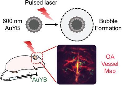
定位光声断层成像(LOT)是最近出现的一种变革性超分辨率技术,它通过对血液中的微粒进行单独定位和跟踪,突破了深部组织光声(OA)成像的声衍射极限。然而,由于红细胞对光的强烈吸收,以前的单颗粒 OA 检测仅限于直径≈5 微米的相对较大的微颗粒。本文证明,直径≈600 nm 的亚微米级多孔金纳米粒子可以单独检测,从而利用 LOT 进行非侵入式超分辨率成像。超高速明场显微镜显示,这些纳米粒子能产生微观等离子气泡,通过纳米到微米的尺寸转换,显著增强光声能量转换。全面的体外和体内测试进一步证明了颗粒的生物相容性和生物安全性。通过将可检测到的颗粒尺寸缩小一个数量级,nanoLOT 实现了微血管成像,大大降低了颗粒聚集造成栓塞的风险,为观察纳米颗粒如何到达血管和潜在的血管外目标开辟了新途径。nanoLOT 在鼠脑微血管网络无创成像方面的性能,有望为神经血管耦合机制以及与神经退行性疾病相关的纵向微循环变化提供新的视角。
Localization optoacoustic tomography (LOT) has recently emerged as a transformative super-resolution technique breaking through the acoustic diffraction limit in deep-tissue optoacoustic (OA) imaging via individual localization and tracking of particles in the bloodstream. However, strong light absorption in red blood cells has previously restricted per-particle OA detection to relatively large microparticles, ≈5 µm in diameter. Herein, it is demonstrated that submicron-sized porous gold nanoparticles, ≈600 nm in diameter, can be individually detected for noninvasive super-resolution imaging with LOT. Ultra-high-speed bright-field microscopy revealed that these nanoparticles generate microscopic plasmonic vapor bubbles, significantly enhancing opto-acoustic energy conversion through a nano-to-micro size transformation. Comprehensive in vitro and in vivo tests further demonstrated the biocompatibility and biosafety of the particles. By reducing the detectable particle size by an order of magnitude, nanoLOT enables microangiographic imaging with a significantly reduced risk of embolisms from particle aggregation and opens new avenues to visualize how nanoparticles reach vascular and potentially extravascular targets. The performance of nanoLOT for non-invasive imaging of microvascular networks in the murine brain anticipates new insights into neurovascular coupling mechanisms and longitudinal microcirculatory changes associated with neurodegenerative diseases.
期刊介绍:
Small serves as an exceptional platform for both experimental and theoretical studies in fundamental and applied interdisciplinary research at the nano- and microscale. The journal offers a compelling mix of peer-reviewed Research Articles, Reviews, Perspectives, and Comments.
With a remarkable 2022 Journal Impact Factor of 13.3 (Journal Citation Reports from Clarivate Analytics, 2023), Small remains among the top multidisciplinary journals, covering a wide range of topics at the interface of materials science, chemistry, physics, engineering, medicine, and biology.
Small's readership includes biochemists, biologists, biomedical scientists, chemists, engineers, information technologists, materials scientists, physicists, and theoreticians alike.


 分享
分享
 求助内容:
求助内容: 应助结果提醒方式:
 扫码关注我们
