Danilo Tadao Wada, Li Siyuan Wada, Camila Vilas Boas Machado, Mateus Repolês Lourenço, Tales Rubens de Nadai, Federico Enrique Garcia Cipriano, Alexandre Todorovic Fabro, Marcel Koenigkam-Santos
{"title":"在肺部病变评估中使用 Look-Locker T1 弛豫测量法和高分辨率 T2:一项单中心前瞻性研究。","authors":"Danilo Tadao Wada, Li Siyuan Wada, Camila Vilas Boas Machado, Mateus Repolês Lourenço, Tales Rubens de Nadai, Federico Enrique Garcia Cipriano, Alexandre Todorovic Fabro, Marcel Koenigkam-Santos","doi":"10.1590/0100-3984.2024.0033","DOIUrl":null,"url":null,"abstract":"<p><strong>Objective: </strong>To explore the feasibility of two magnetic resonance imaging (MRI) sequences-high-resolution T2-weighted (HR T2) and Look-Locker T1 (LL T1) relaxometry-for the investigation focal lung lesions (FLLs). As a secondary objective, we analyzed the diagnostic accuracy of these sequences.</p><p><strong>Materials and methods: </strong>This was a prospective observational study involving 39 subjects with FLLs scanned in a 1.5-T MRI system with LL T1 relaxometry and HR T2 sequences focused on the FLL region, in addition to a conventional protocol. All images were evaluated by two radiologists, working independently, who were blinded to other findings.</p><p><strong>Results: </strong>Most of the examinations (31 of the LL T1 relaxometry sequences and 36 of the HR T2 sequences) were of adequate diagnostic quality. Nondiagnostic examinations were considered so mainly because of limited coverage of the sequences. Of the FLLs studied, 19 were malignant, 17 were benign, and three were excluded from the accuracy analysis because there was no definitive diagnosis. Although LL T1 relaxometry could not distinguish between benign and malignant lesions, the signal intensity at its first inversion time (160 ms) differed between the two groups. The HR T2 sequence was considered the best sequence for assessing specific morphological characteristics, especially pseudocavities and pleural tags. We found that MRI showed better accuracy than did computed tomography (86% vs. 74%).</p><p><strong>Conclusion: </strong>Both MRI sequences are feasible for the evaluation of FLLs. Images at 160 ms of the LL T1 relaxometry sequence helped distinguish between benign and malignant lesions, and the HR T2 sequence was considered the best sequence for evaluating specific morphological characteristics.</p>","PeriodicalId":20842,"journal":{"name":"Radiologia Brasileira","volume":"57 ","pages":"e20240033"},"PeriodicalIF":0.0000,"publicationDate":"2024-09-30","publicationTypes":"Journal Article","fieldsOfStudy":null,"isOpenAccess":false,"openAccessPdf":"https://www.ncbi.nlm.nih.gov/pmc/articles/PMC11469640/pdf/","citationCount":"0","resultStr":"{\"title\":\"Look-Locker T1 relaxometry and high-resolution T2 in the evaluation of lung lesions: a single-center prospective study.\",\"authors\":\"Danilo Tadao Wada, Li Siyuan Wada, Camila Vilas Boas Machado, Mateus Repolês Lourenço, Tales Rubens de Nadai, Federico Enrique Garcia Cipriano, Alexandre Todorovic Fabro, Marcel Koenigkam-Santos\",\"doi\":\"10.1590/0100-3984.2024.0033\",\"DOIUrl\":null,\"url\":null,\"abstract\":\"<p><strong>Objective: </strong>To explore the feasibility of two magnetic resonance imaging (MRI) sequences-high-resolution T2-weighted (HR T2) and Look-Locker T1 (LL T1) relaxometry-for the investigation focal lung lesions (FLLs). As a secondary objective, we analyzed the diagnostic accuracy of these sequences.</p><p><strong>Materials and methods: </strong>This was a prospective observational study involving 39 subjects with FLLs scanned in a 1.5-T MRI system with LL T1 relaxometry and HR T2 sequences focused on the FLL region, in addition to a conventional protocol. All images were evaluated by two radiologists, working independently, who were blinded to other findings.</p><p><strong>Results: </strong>Most of the examinations (31 of the LL T1 relaxometry sequences and 36 of the HR T2 sequences) were of adequate diagnostic quality. Nondiagnostic examinations were considered so mainly because of limited coverage of the sequences. Of the FLLs studied, 19 were malignant, 17 were benign, and three were excluded from the accuracy analysis because there was no definitive diagnosis. Although LL T1 relaxometry could not distinguish between benign and malignant lesions, the signal intensity at its first inversion time (160 ms) differed between the two groups. The HR T2 sequence was considered the best sequence for assessing specific morphological characteristics, especially pseudocavities and pleural tags. We found that MRI showed better accuracy than did computed tomography (86% vs. 74%).</p><p><strong>Conclusion: </strong>Both MRI sequences are feasible for the evaluation of FLLs. Images at 160 ms of the LL T1 relaxometry sequence helped distinguish between benign and malignant lesions, and the HR T2 sequence was considered the best sequence for evaluating specific morphological characteristics.</p>\",\"PeriodicalId\":20842,\"journal\":{\"name\":\"Radiologia Brasileira\",\"volume\":\"57 \",\"pages\":\"e20240033\"},\"PeriodicalIF\":0.0000,\"publicationDate\":\"2024-09-30\",\"publicationTypes\":\"Journal Article\",\"fieldsOfStudy\":null,\"isOpenAccess\":false,\"openAccessPdf\":\"https://www.ncbi.nlm.nih.gov/pmc/articles/PMC11469640/pdf/\",\"citationCount\":\"0\",\"resultStr\":null,\"platform\":\"Semanticscholar\",\"paperid\":null,\"PeriodicalName\":\"Radiologia Brasileira\",\"FirstCategoryId\":\"1085\",\"ListUrlMain\":\"https://doi.org/10.1590/0100-3984.2024.0033\",\"RegionNum\":0,\"RegionCategory\":null,\"ArticlePicture\":[],\"TitleCN\":null,\"AbstractTextCN\":null,\"PMCID\":null,\"EPubDate\":\"2024/1/1 0:00:00\",\"PubModel\":\"eCollection\",\"JCR\":\"Q3\",\"JCRName\":\"Medicine\",\"Score\":null,\"Total\":0}","platform":"Semanticscholar","paperid":null,"PeriodicalName":"Radiologia Brasileira","FirstCategoryId":"1085","ListUrlMain":"https://doi.org/10.1590/0100-3984.2024.0033","RegionNum":0,"RegionCategory":null,"ArticlePicture":[],"TitleCN":null,"AbstractTextCN":null,"PMCID":null,"EPubDate":"2024/1/1 0:00:00","PubModel":"eCollection","JCR":"Q3","JCRName":"Medicine","Score":null,"Total":0}
Look-Locker T1 relaxometry and high-resolution T2 in the evaluation of lung lesions: a single-center prospective study.
Objective: To explore the feasibility of two magnetic resonance imaging (MRI) sequences-high-resolution T2-weighted (HR T2) and Look-Locker T1 (LL T1) relaxometry-for the investigation focal lung lesions (FLLs). As a secondary objective, we analyzed the diagnostic accuracy of these sequences.
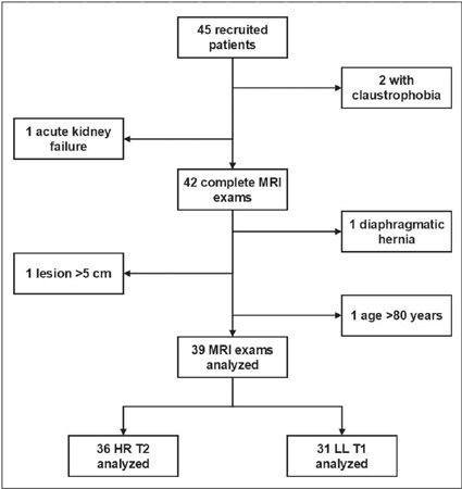
Materials and methods: This was a prospective observational study involving 39 subjects with FLLs scanned in a 1.5-T MRI system with LL T1 relaxometry and HR T2 sequences focused on the FLL region, in addition to a conventional protocol. All images were evaluated by two radiologists, working independently, who were blinded to other findings.
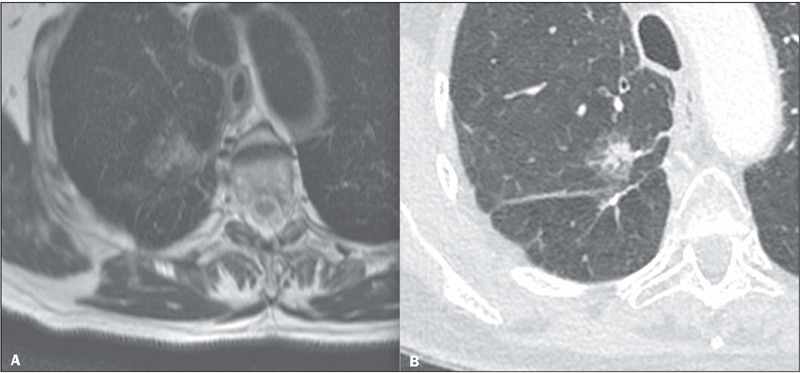
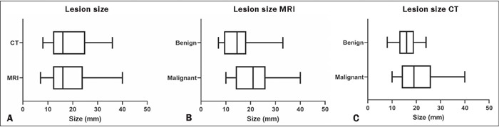
Results: Most of the examinations (31 of the LL T1 relaxometry sequences and 36 of the HR T2 sequences) were of adequate diagnostic quality. Nondiagnostic examinations were considered so mainly because of limited coverage of the sequences. Of the FLLs studied, 19 were malignant, 17 were benign, and three were excluded from the accuracy analysis because there was no definitive diagnosis. Although LL T1 relaxometry could not distinguish between benign and malignant lesions, the signal intensity at its first inversion time (160 ms) differed between the two groups. The HR T2 sequence was considered the best sequence for assessing specific morphological characteristics, especially pseudocavities and pleural tags. We found that MRI showed better accuracy than did computed tomography (86% vs. 74%).
Conclusion: Both MRI sequences are feasible for the evaluation of FLLs. Images at 160 ms of the LL T1 relaxometry sequence helped distinguish between benign and malignant lesions, and the HR T2 sequence was considered the best sequence for evaluating specific morphological characteristics.


 分享
分享
 求助内容:
求助内容: 应助结果提醒方式:
 扫码关注我们
