{"title":"核磁共振成像显示黄粒肉芽肿性前列腺炎伴多发性肿块。","authors":"Takashi Okamoto, Kazuki Doi, Soma Ogura, Masafumi Kusunose, Kosuke Takahashi, Takuya Fujimoto, Mototsugu Muramaki, Yuji Yamada","doi":"10.1002/iju5.12791","DOIUrl":null,"url":null,"abstract":"<div>\n \n <section>\n \n <h3> Introduction</h3>\n \n <p>Xanthogranulomatous prostatitis is a very rare benign inflammatory lesion of the prostate that may be similar to prostatic carcinoma in clinical presentation and radiological characteristics.</p>\n </section>\n \n <section>\n \n <h3> Case presentation</h3>\n \n <p>A 77-year-old man was admitted to our hospital because of high prostate-specific antigen level. Magnetic resonance imaging showed a 6.5 cm in diameter multifocal mass with hemorrhage at the base of the left lobe of the prostate. Biopsy was performed. Histopathological examination revealed no evidence of malignancy. After biopsy, he developed recurring fever, so the patient was treated with open suprapubic tumor resection to control infection. Pathological examination revealed xanthogranulomatous prostatitis.</p>\n </section>\n \n <section>\n \n <h3> Conclusion</h3>\n \n <p>It is necessary to diagnose xanthogranulomatous prostatitis by cooperation between urologists and pathologists, and consider xanthogranulomatous prostatitis as a differential diagnosis. Treatment should be conservative in principle; however, surgical intervention may be necessary.</p>\n </section>\n </div>","PeriodicalId":52909,"journal":{"name":"IJU Case Reports","volume":"7 6","pages":"503-505"},"PeriodicalIF":0.5000,"publicationDate":"2024-09-26","publicationTypes":"Journal Article","fieldsOfStudy":null,"isOpenAccess":false,"openAccessPdf":"https://www.ncbi.nlm.nih.gov/pmc/articles/PMC11531873/pdf/","citationCount":"0","resultStr":"{\"title\":\"Xanthogranulomatous prostatitis with multilocular mass on MRI\",\"authors\":\"Takashi Okamoto, Kazuki Doi, Soma Ogura, Masafumi Kusunose, Kosuke Takahashi, Takuya Fujimoto, Mototsugu Muramaki, Yuji Yamada\",\"doi\":\"10.1002/iju5.12791\",\"DOIUrl\":null,\"url\":null,\"abstract\":\"<div>\\n \\n <section>\\n \\n <h3> Introduction</h3>\\n \\n <p>Xanthogranulomatous prostatitis is a very rare benign inflammatory lesion of the prostate that may be similar to prostatic carcinoma in clinical presentation and radiological characteristics.</p>\\n </section>\\n \\n <section>\\n \\n <h3> Case presentation</h3>\\n \\n <p>A 77-year-old man was admitted to our hospital because of high prostate-specific antigen level. Magnetic resonance imaging showed a 6.5 cm in diameter multifocal mass with hemorrhage at the base of the left lobe of the prostate. Biopsy was performed. Histopathological examination revealed no evidence of malignancy. After biopsy, he developed recurring fever, so the patient was treated with open suprapubic tumor resection to control infection. Pathological examination revealed xanthogranulomatous prostatitis.</p>\\n </section>\\n \\n <section>\\n \\n <h3> Conclusion</h3>\\n \\n <p>It is necessary to diagnose xanthogranulomatous prostatitis by cooperation between urologists and pathologists, and consider xanthogranulomatous prostatitis as a differential diagnosis. Treatment should be conservative in principle; however, surgical intervention may be necessary.</p>\\n </section>\\n </div>\",\"PeriodicalId\":52909,\"journal\":{\"name\":\"IJU Case Reports\",\"volume\":\"7 6\",\"pages\":\"503-505\"},\"PeriodicalIF\":0.5000,\"publicationDate\":\"2024-09-26\",\"publicationTypes\":\"Journal Article\",\"fieldsOfStudy\":null,\"isOpenAccess\":false,\"openAccessPdf\":\"https://www.ncbi.nlm.nih.gov/pmc/articles/PMC11531873/pdf/\",\"citationCount\":\"0\",\"resultStr\":null,\"platform\":\"Semanticscholar\",\"paperid\":null,\"PeriodicalName\":\"IJU Case Reports\",\"FirstCategoryId\":\"1085\",\"ListUrlMain\":\"https://onlinelibrary.wiley.com/doi/10.1002/iju5.12791\",\"RegionNum\":0,\"RegionCategory\":null,\"ArticlePicture\":[],\"TitleCN\":null,\"AbstractTextCN\":null,\"PMCID\":null,\"EPubDate\":\"\",\"PubModel\":\"\",\"JCR\":\"Q4\",\"JCRName\":\"Medicine\",\"Score\":null,\"Total\":0}","platform":"Semanticscholar","paperid":null,"PeriodicalName":"IJU Case Reports","FirstCategoryId":"1085","ListUrlMain":"https://onlinelibrary.wiley.com/doi/10.1002/iju5.12791","RegionNum":0,"RegionCategory":null,"ArticlePicture":[],"TitleCN":null,"AbstractTextCN":null,"PMCID":null,"EPubDate":"","PubModel":"","JCR":"Q4","JCRName":"Medicine","Score":null,"Total":0}
Xanthogranulomatous prostatitis with multilocular mass on MRI
Introduction
Xanthogranulomatous prostatitis is a very rare benign inflammatory lesion of the prostate that may be similar to prostatic carcinoma in clinical presentation and radiological characteristics.
Case presentation
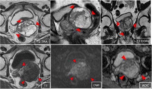
A 77-year-old man was admitted to our hospital because of high prostate-specific antigen level. Magnetic resonance imaging showed a 6.5 cm in diameter multifocal mass with hemorrhage at the base of the left lobe of the prostate. Biopsy was performed. Histopathological examination revealed no evidence of malignancy. After biopsy, he developed recurring fever, so the patient was treated with open suprapubic tumor resection to control infection. Pathological examination revealed xanthogranulomatous prostatitis.
Conclusion
It is necessary to diagnose xanthogranulomatous prostatitis by cooperation between urologists and pathologists, and consider xanthogranulomatous prostatitis as a differential diagnosis. Treatment should be conservative in principle; however, surgical intervention may be necessary.


 分享
分享
 求助内容:
求助内容: 应助结果提醒方式:
 扫码关注我们
