Kostas G. Mavrakis, Gerasimos Divaris, Maria Tampakaki, Saba N. Khan, Kishan Dholakia, Giannis Zacharakis
{"title":"光学生成的液滴束改善了作为阿尔茨海默病生物标志物的脉络膜厚度的光声成像效果","authors":"Kostas G. Mavrakis, Gerasimos Divaris, Maria Tampakaki, Saba N. Khan, Kishan Dholakia, Giannis Zacharakis","doi":"10.1038/s44310-024-00036-3","DOIUrl":null,"url":null,"abstract":"Optoacoustic microscopy faces a restricted depth of field attributed to the tightly focused Gaussian beam excitation. This limitation poses challenges in capturing high-resolution images of samples with uneven surfaces or obtaining high-quality volumetric images without z-scanning. To address this issue, we propose the use of droplet beam illumination in optoacoustic microscopy, which extends the depth of field to approximately 80 times the Rayleigh length. The droplet beam is generated using a Mach–Zehnder-type interferometer, with each arm equipped with a lens of different optical power. We demonstrate the advantages of droplet beam illumination in microscopy by showing high contrast images on fluorescent beads with a 50% improvement compared to Bessel beam illumination and subsequently imaging the posterior cavity of mice eyes. This method introduces novel perspectives to medical sciences, allowing the measurement of the choroidal layer thickness, an early indicative biomarker for Alzheimer’s disease.","PeriodicalId":501711,"journal":{"name":"npj Nanophotonics","volume":" ","pages":"1-9"},"PeriodicalIF":0.0000,"publicationDate":"2024-11-05","publicationTypes":"Journal Article","fieldsOfStudy":null,"isOpenAccess":false,"openAccessPdf":"https://www.nature.com/articles/s44310-024-00036-3.pdf","citationCount":"0","resultStr":"{\"title\":\"Optically generated droplet beams improve optoacoustic imaging of choroid thickness as an Alzheimer’s disease biomarker\",\"authors\":\"Kostas G. Mavrakis, Gerasimos Divaris, Maria Tampakaki, Saba N. Khan, Kishan Dholakia, Giannis Zacharakis\",\"doi\":\"10.1038/s44310-024-00036-3\",\"DOIUrl\":null,\"url\":null,\"abstract\":\"Optoacoustic microscopy faces a restricted depth of field attributed to the tightly focused Gaussian beam excitation. This limitation poses challenges in capturing high-resolution images of samples with uneven surfaces or obtaining high-quality volumetric images without z-scanning. To address this issue, we propose the use of droplet beam illumination in optoacoustic microscopy, which extends the depth of field to approximately 80 times the Rayleigh length. The droplet beam is generated using a Mach–Zehnder-type interferometer, with each arm equipped with a lens of different optical power. We demonstrate the advantages of droplet beam illumination in microscopy by showing high contrast images on fluorescent beads with a 50% improvement compared to Bessel beam illumination and subsequently imaging the posterior cavity of mice eyes. This method introduces novel perspectives to medical sciences, allowing the measurement of the choroidal layer thickness, an early indicative biomarker for Alzheimer’s disease.\",\"PeriodicalId\":501711,\"journal\":{\"name\":\"npj Nanophotonics\",\"volume\":\" \",\"pages\":\"1-9\"},\"PeriodicalIF\":0.0000,\"publicationDate\":\"2024-11-05\",\"publicationTypes\":\"Journal Article\",\"fieldsOfStudy\":null,\"isOpenAccess\":false,\"openAccessPdf\":\"https://www.nature.com/articles/s44310-024-00036-3.pdf\",\"citationCount\":\"0\",\"resultStr\":null,\"platform\":\"Semanticscholar\",\"paperid\":null,\"PeriodicalName\":\"npj Nanophotonics\",\"FirstCategoryId\":\"1085\",\"ListUrlMain\":\"https://www.nature.com/articles/s44310-024-00036-3\",\"RegionNum\":0,\"RegionCategory\":null,\"ArticlePicture\":[],\"TitleCN\":null,\"AbstractTextCN\":null,\"PMCID\":null,\"EPubDate\":\"\",\"PubModel\":\"\",\"JCR\":\"\",\"JCRName\":\"\",\"Score\":null,\"Total\":0}","platform":"Semanticscholar","paperid":null,"PeriodicalName":"npj Nanophotonics","FirstCategoryId":"1085","ListUrlMain":"https://www.nature.com/articles/s44310-024-00036-3","RegionNum":0,"RegionCategory":null,"ArticlePicture":[],"TitleCN":null,"AbstractTextCN":null,"PMCID":null,"EPubDate":"","PubModel":"","JCR":"","JCRName":"","Score":null,"Total":0}
引用次数: 0
摘要
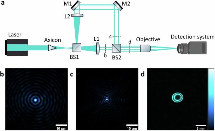
光声显微镜面临着景深受限的问题,这是由于紧聚焦高斯光束激发的缘故。这种限制给捕捉表面凹凸不平的样品的高分辨率图像或在不进行 Z 扫描的情况下获得高质量的体积图像带来了挑战。为了解决这个问题,我们提出在光声显微镜中使用液滴光束照明,它能将景深扩展到约 80 倍的瑞利长度。液滴光束是利用马赫-泽恩德型干涉仪产生的,每个臂都配备了不同光功率的透镜。我们展示了液滴光束照明在显微镜中的优势,与贝塞尔光束照明相比,荧光珠上的高对比度图像提高了 50%,随后还对小鼠眼球后腔进行了成像。这种方法为医学科学引入了新的视角,可以测量脉络膜层厚度,这是阿尔茨海默病的早期指示性生物标志物。
Optically generated droplet beams improve optoacoustic imaging of choroid thickness as an Alzheimer’s disease biomarker
Optoacoustic microscopy faces a restricted depth of field attributed to the tightly focused Gaussian beam excitation. This limitation poses challenges in capturing high-resolution images of samples with uneven surfaces or obtaining high-quality volumetric images without z-scanning. To address this issue, we propose the use of droplet beam illumination in optoacoustic microscopy, which extends the depth of field to approximately 80 times the Rayleigh length. The droplet beam is generated using a Mach–Zehnder-type interferometer, with each arm equipped with a lens of different optical power. We demonstrate the advantages of droplet beam illumination in microscopy by showing high contrast images on fluorescent beads with a 50% improvement compared to Bessel beam illumination and subsequently imaging the posterior cavity of mice eyes. This method introduces novel perspectives to medical sciences, allowing the measurement of the choroidal layer thickness, an early indicative biomarker for Alzheimer’s disease.


 分享
分享

 求助内容:
求助内容: 应助结果提醒方式:
 扫码关注我们
