Boyang Mao , Hong Wang , Hongxi Zhang , Xueliang Shang , Zhi Yang
{"title":"学龄前儿童胼胝体厚度的发展趋势","authors":"Boyang Mao , Hong Wang , Hongxi Zhang , Xueliang Shang , Zhi Yang","doi":"10.1016/j.metrad.2024.100111","DOIUrl":null,"url":null,"abstract":"<div><h3>Background</h3><div>The corpus callosum plays a crucial role in integrated brain functions, and its development in childhood is strongly associated with subsequent cognitive, emotional, and behavioral development. However, there is still a lack of clear understanding regarding the developmental trends of the corpus callosum in preschool children. This study aims to comprehensively investigate age and sex differences in the thickness of the corpus callosum in typical developing children between 1 and 6 years old.</div></div><div><h3>Methods</h3><div>T1-weighted structural MRI data were collected from a sample of 295 neurologically normal children aged 1–6 years. Utilizing the specialized corpus callosum segmentation software Yuki, thickness measurements of the mid-sagittal plane of the corpus callosum were obtained.</div></div><div><h3>Results</h3><div>The anterior part exhibited faster growth compared to the middle and posterior sections, while growth at the extremities was not statistically significant. Furthermore, gender differences were identified, with males showing earlier development of the corpus callosum, particularly between ages 1 and 3. Conversely, females exhibited the most notable increase in thickness between ages 3 and 5.</div></div><div><h3>Conclusion</h3><div>This study provides significant insights into the developmental trends of the mid-sagittal plane of the corpus callosum in preschool children. It reveals distinct non-linear developmental patterns in different sections of the corpus callosum and highlights the influence of sex on these developmental patterns.</div></div>","PeriodicalId":100921,"journal":{"name":"Meta-Radiology","volume":"2 4","pages":"Article 100111"},"PeriodicalIF":0.0000,"publicationDate":"2024-12-01","publicationTypes":"Journal Article","fieldsOfStudy":null,"isOpenAccess":false,"openAccessPdf":"","citationCount":"0","resultStr":"{\"title\":\"Developmental trends in corpus callosum thickness among preschool children\",\"authors\":\"Boyang Mao , Hong Wang , Hongxi Zhang , Xueliang Shang , Zhi Yang\",\"doi\":\"10.1016/j.metrad.2024.100111\",\"DOIUrl\":null,\"url\":null,\"abstract\":\"<div><h3>Background</h3><div>The corpus callosum plays a crucial role in integrated brain functions, and its development in childhood is strongly associated with subsequent cognitive, emotional, and behavioral development. However, there is still a lack of clear understanding regarding the developmental trends of the corpus callosum in preschool children. This study aims to comprehensively investigate age and sex differences in the thickness of the corpus callosum in typical developing children between 1 and 6 years old.</div></div><div><h3>Methods</h3><div>T1-weighted structural MRI data were collected from a sample of 295 neurologically normal children aged 1–6 years. Utilizing the specialized corpus callosum segmentation software Yuki, thickness measurements of the mid-sagittal plane of the corpus callosum were obtained.</div></div><div><h3>Results</h3><div>The anterior part exhibited faster growth compared to the middle and posterior sections, while growth at the extremities was not statistically significant. Furthermore, gender differences were identified, with males showing earlier development of the corpus callosum, particularly between ages 1 and 3. Conversely, females exhibited the most notable increase in thickness between ages 3 and 5.</div></div><div><h3>Conclusion</h3><div>This study provides significant insights into the developmental trends of the mid-sagittal plane of the corpus callosum in preschool children. It reveals distinct non-linear developmental patterns in different sections of the corpus callosum and highlights the influence of sex on these developmental patterns.</div></div>\",\"PeriodicalId\":100921,\"journal\":{\"name\":\"Meta-Radiology\",\"volume\":\"2 4\",\"pages\":\"Article 100111\"},\"PeriodicalIF\":0.0000,\"publicationDate\":\"2024-12-01\",\"publicationTypes\":\"Journal Article\",\"fieldsOfStudy\":null,\"isOpenAccess\":false,\"openAccessPdf\":\"\",\"citationCount\":\"0\",\"resultStr\":null,\"platform\":\"Semanticscholar\",\"paperid\":null,\"PeriodicalName\":\"Meta-Radiology\",\"FirstCategoryId\":\"1085\",\"ListUrlMain\":\"https://www.sciencedirect.com/science/article/pii/S2950162824000651\",\"RegionNum\":0,\"RegionCategory\":null,\"ArticlePicture\":[],\"TitleCN\":null,\"AbstractTextCN\":null,\"PMCID\":null,\"EPubDate\":\"2024/10/1 0:00:00\",\"PubModel\":\"Epub\",\"JCR\":\"\",\"JCRName\":\"\",\"Score\":null,\"Total\":0}","platform":"Semanticscholar","paperid":null,"PeriodicalName":"Meta-Radiology","FirstCategoryId":"1085","ListUrlMain":"https://www.sciencedirect.com/science/article/pii/S2950162824000651","RegionNum":0,"RegionCategory":null,"ArticlePicture":[],"TitleCN":null,"AbstractTextCN":null,"PMCID":null,"EPubDate":"2024/10/1 0:00:00","PubModel":"Epub","JCR":"","JCRName":"","Score":null,"Total":0}
Developmental trends in corpus callosum thickness among preschool children
Background
The corpus callosum plays a crucial role in integrated brain functions, and its development in childhood is strongly associated with subsequent cognitive, emotional, and behavioral development. However, there is still a lack of clear understanding regarding the developmental trends of the corpus callosum in preschool children. This study aims to comprehensively investigate age and sex differences in the thickness of the corpus callosum in typical developing children between 1 and 6 years old.
Methods
T1-weighted structural MRI data were collected from a sample of 295 neurologically normal children aged 1–6 years. Utilizing the specialized corpus callosum segmentation software Yuki, thickness measurements of the mid-sagittal plane of the corpus callosum were obtained.
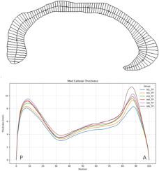
Results
The anterior part exhibited faster growth compared to the middle and posterior sections, while growth at the extremities was not statistically significant. Furthermore, gender differences were identified, with males showing earlier development of the corpus callosum, particularly between ages 1 and 3. Conversely, females exhibited the most notable increase in thickness between ages 3 and 5.
Conclusion
This study provides significant insights into the developmental trends of the mid-sagittal plane of the corpus callosum in preschool children. It reveals distinct non-linear developmental patterns in different sections of the corpus callosum and highlights the influence of sex on these developmental patterns.


 分享
分享

 求助内容:
求助内容: 应助结果提醒方式:
 扫码关注我们
