Alzbeta Dikunova, Nikola Noskova, Jan H. Overbeck, Martin Polak, David Stelzig, David Zapletal, Karel Kubicek, Jiri Novacek, Remco Sprangers, Richard Stefl
{"title":"中温和亲热生物中Xrn2/ Rat1-Rai1-Rtt103末端复合物的组装","authors":"Alzbeta Dikunova, Nikola Noskova, Jan H. Overbeck, Martin Polak, David Stelzig, David Zapletal, Karel Kubicek, Jiri Novacek, Remco Sprangers, Richard Stefl","doi":"10.1016/j.str.2024.11.010","DOIUrl":null,"url":null,"abstract":"The 5′–3′ exoribonuclease Xrn2, known as Rat1 in yeasts, terminates mRNA transcription by RNA polymerase II (RNAPII). In the torpedo model of termination, the activity of Xrn2/Rat1 is enhanced by Rai1, which is recruited to the termination site by Rtt103, an adaptor protein binding to the RNAPII C-terminal domain (CTD). The overall architecture of the Xrn2/Rat1-Rai1-Rtt103 complex remains unknown. We combined structural biology methods to characterize the torpedo complex from <em>Saccharomyces cerevisiae</em> and <em>Chaetomium thermophilum</em>. Comparison of the structures from these organisms revealed a conserved protein core fold of the subunits, but significant variability in their interaction interfaces. We found that in the mesophile, Rtt103 utilizes an unstructured region to augment a Rai1 β-sheet, while in the thermophile Rtt103 binds to a C-terminal helix of Rai1 via its CTD-interacting domain with an α-helical fold. These different torpedo complex assemblies reflect adaptations to the environment and impact complex recruitment to RNAPII.","PeriodicalId":22168,"journal":{"name":"Structure","volume":"34 1","pages":""},"PeriodicalIF":4.4000,"publicationDate":"2024-12-09","publicationTypes":"Journal Article","fieldsOfStudy":null,"isOpenAccess":false,"openAccessPdf":"","citationCount":"0","resultStr":"{\"title\":\"Assembly of the Xrn2/Rat1–Rai1–Rtt103 termination complexes in mesophilic and thermophilic organisms\",\"authors\":\"Alzbeta Dikunova, Nikola Noskova, Jan H. Overbeck, Martin Polak, David Stelzig, David Zapletal, Karel Kubicek, Jiri Novacek, Remco Sprangers, Richard Stefl\",\"doi\":\"10.1016/j.str.2024.11.010\",\"DOIUrl\":null,\"url\":null,\"abstract\":\"The 5′–3′ exoribonuclease Xrn2, known as Rat1 in yeasts, terminates mRNA transcription by RNA polymerase II (RNAPII). In the torpedo model of termination, the activity of Xrn2/Rat1 is enhanced by Rai1, which is recruited to the termination site by Rtt103, an adaptor protein binding to the RNAPII C-terminal domain (CTD). The overall architecture of the Xrn2/Rat1-Rai1-Rtt103 complex remains unknown. We combined structural biology methods to characterize the torpedo complex from <em>Saccharomyces cerevisiae</em> and <em>Chaetomium thermophilum</em>. Comparison of the structures from these organisms revealed a conserved protein core fold of the subunits, but significant variability in their interaction interfaces. We found that in the mesophile, Rtt103 utilizes an unstructured region to augment a Rai1 β-sheet, while in the thermophile Rtt103 binds to a C-terminal helix of Rai1 via its CTD-interacting domain with an α-helical fold. These different torpedo complex assemblies reflect adaptations to the environment and impact complex recruitment to RNAPII.\",\"PeriodicalId\":22168,\"journal\":{\"name\":\"Structure\",\"volume\":\"34 1\",\"pages\":\"\"},\"PeriodicalIF\":4.4000,\"publicationDate\":\"2024-12-09\",\"publicationTypes\":\"Journal Article\",\"fieldsOfStudy\":null,\"isOpenAccess\":false,\"openAccessPdf\":\"\",\"citationCount\":\"0\",\"resultStr\":null,\"platform\":\"Semanticscholar\",\"paperid\":null,\"PeriodicalName\":\"Structure\",\"FirstCategoryId\":\"99\",\"ListUrlMain\":\"https://doi.org/10.1016/j.str.2024.11.010\",\"RegionNum\":2,\"RegionCategory\":\"生物学\",\"ArticlePicture\":[],\"TitleCN\":null,\"AbstractTextCN\":null,\"PMCID\":null,\"EPubDate\":\"\",\"PubModel\":\"\",\"JCR\":\"Q2\",\"JCRName\":\"BIOCHEMISTRY & MOLECULAR BIOLOGY\",\"Score\":null,\"Total\":0}","platform":"Semanticscholar","paperid":null,"PeriodicalName":"Structure","FirstCategoryId":"99","ListUrlMain":"https://doi.org/10.1016/j.str.2024.11.010","RegionNum":2,"RegionCategory":"生物学","ArticlePicture":[],"TitleCN":null,"AbstractTextCN":null,"PMCID":null,"EPubDate":"","PubModel":"","JCR":"Q2","JCRName":"BIOCHEMISTRY & MOLECULAR BIOLOGY","Score":null,"Total":0}
引用次数: 0
摘要
5 ‘ -3 ’外核糖核酸酶Xrn2,在酵母中被称为Rat1,通过RNA聚合酶II (RNAPII)终止mRNA转录。在鱼雷终止模型中,Rai1增强了Xrn2/Rat1的活性,Rai1通过Rtt103招募到终止位点,Rtt103是一种结合RNAPII c -末端结构域(CTD)的接头蛋白。Xrn2/Rat1-Rai1-Rtt103复合体的整体结构仍然未知。我们结合结构生物学方法对酿酒酵母和嗜热毛菌的鱼雷复合物进行了表征。这些生物的结构比较揭示了亚基的保守蛋白核心折叠,但它们的相互作用界面有显著的差异。我们发现,在亲介菌中,Rtt103利用一个非结构化区域来增加Rai1 β-片,而在亲热菌中,Rtt103通过其具有α-螺旋折叠的ctd相互作用结构域与Rai1的c端螺旋结合。这些不同的鱼雷复合物装配反映了对环境的适应和影响RNAPII的复合物招募。
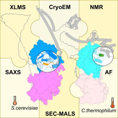
Assembly of the Xrn2/Rat1–Rai1–Rtt103 termination complexes in mesophilic and thermophilic organisms
The 5′–3′ exoribonuclease Xrn2, known as Rat1 in yeasts, terminates mRNA transcription by RNA polymerase II (RNAPII). In the torpedo model of termination, the activity of Xrn2/Rat1 is enhanced by Rai1, which is recruited to the termination site by Rtt103, an adaptor protein binding to the RNAPII C-terminal domain (CTD). The overall architecture of the Xrn2/Rat1-Rai1-Rtt103 complex remains unknown. We combined structural biology methods to characterize the torpedo complex from Saccharomyces cerevisiae and Chaetomium thermophilum. Comparison of the structures from these organisms revealed a conserved protein core fold of the subunits, but significant variability in their interaction interfaces. We found that in the mesophile, Rtt103 utilizes an unstructured region to augment a Rai1 β-sheet, while in the thermophile Rtt103 binds to a C-terminal helix of Rai1 via its CTD-interacting domain with an α-helical fold. These different torpedo complex assemblies reflect adaptations to the environment and impact complex recruitment to RNAPII.
期刊介绍:
Structure aims to publish papers of exceptional interest in the field of structural biology. The journal strives to be essential reading for structural biologists, as well as biologists and biochemists that are interested in macromolecular structure and function. Structure strongly encourages the submission of manuscripts that present structural and molecular insights into biological function and mechanism. Other reports that address fundamental questions in structural biology, such as structure-based examinations of protein evolution, folding, and/or design, will also be considered. We will consider the application of any method, experimental or computational, at high or low resolution, to conduct structural investigations, as long as the method is appropriate for the biological, functional, and mechanistic question(s) being addressed. Likewise, reports describing single-molecule analysis of biological mechanisms are welcome.
In general, the editors encourage submission of experimental structural studies that are enriched by an analysis of structure-activity relationships and will not consider studies that solely report structural information unless the structure or analysis is of exceptional and broad interest. Studies reporting only homology models, de novo models, or molecular dynamics simulations are also discouraged unless the models are informed by or validated by novel experimental data; rationalization of a large body of existing experimental evidence and making testable predictions based on a model or simulation is often not considered sufficient.


 分享
分享
 求助内容:
求助内容: 应助结果提醒方式:
 扫码关注我们
