{"title":"白点综合征多模态影像学研究进展。","authors":"Ahana Sen, Chetan Rao, Jyotirmay Biswas","doi":"10.4103/ojo.ojo_116_24","DOIUrl":null,"url":null,"abstract":"<p><p>The white dot syndromes are a group of phenotypically similar disorders characterized by multiple lesions at the level of the outer retina, retinal pigment epithelium, and choroid. Common white dot syndromes whose imaging modalities have been described in this article are multiple evanescent white dot syndrome, acute posterior multifocal placoid pigment epitheliopathy, acute zonal occult outer retinopathy, multifocal choroiditis and panuveitis, punctate inner choroidopathy, serpiginous choroiditis, and birdshot chorioretinopathy. The various imaging modalities help us to better understand the pathophysiology of the various entities and help in diagnosing, monitoring, and prognosticating them. Optical coherence tomography angiography (OCTA) is a comparatively newer tool that helps us to visualize lesions in the choroid that correlate with indocyanine green angiography (ICGA) findings. Even though it is of limited value and cannot replace ICGA, it had gained considerable interest among ophthalmologists. Similarly, the noninvasive nature of modalities such as fundus autofluorescence and OCT makes them appealing and preferable over invasive techniques such as fundus fluorescein angiography and ICGA.</p>","PeriodicalId":19461,"journal":{"name":"Oman Journal of Ophthalmology","volume":"17 3","pages":"325-333"},"PeriodicalIF":0.0000,"publicationDate":"2024-10-24","publicationTypes":"Journal Article","fieldsOfStudy":null,"isOpenAccess":false,"openAccessPdf":"https://www.ncbi.nlm.nih.gov/pmc/articles/PMC11620295/pdf/","citationCount":"0","resultStr":"{\"title\":\"An update of multimodal imaging in white dot syndrome.\",\"authors\":\"Ahana Sen, Chetan Rao, Jyotirmay Biswas\",\"doi\":\"10.4103/ojo.ojo_116_24\",\"DOIUrl\":null,\"url\":null,\"abstract\":\"<p><p>The white dot syndromes are a group of phenotypically similar disorders characterized by multiple lesions at the level of the outer retina, retinal pigment epithelium, and choroid. Common white dot syndromes whose imaging modalities have been described in this article are multiple evanescent white dot syndrome, acute posterior multifocal placoid pigment epitheliopathy, acute zonal occult outer retinopathy, multifocal choroiditis and panuveitis, punctate inner choroidopathy, serpiginous choroiditis, and birdshot chorioretinopathy. The various imaging modalities help us to better understand the pathophysiology of the various entities and help in diagnosing, monitoring, and prognosticating them. Optical coherence tomography angiography (OCTA) is a comparatively newer tool that helps us to visualize lesions in the choroid that correlate with indocyanine green angiography (ICGA) findings. Even though it is of limited value and cannot replace ICGA, it had gained considerable interest among ophthalmologists. Similarly, the noninvasive nature of modalities such as fundus autofluorescence and OCT makes them appealing and preferable over invasive techniques such as fundus fluorescein angiography and ICGA.</p>\",\"PeriodicalId\":19461,\"journal\":{\"name\":\"Oman Journal of Ophthalmology\",\"volume\":\"17 3\",\"pages\":\"325-333\"},\"PeriodicalIF\":0.0000,\"publicationDate\":\"2024-10-24\",\"publicationTypes\":\"Journal Article\",\"fieldsOfStudy\":null,\"isOpenAccess\":false,\"openAccessPdf\":\"https://www.ncbi.nlm.nih.gov/pmc/articles/PMC11620295/pdf/\",\"citationCount\":\"0\",\"resultStr\":null,\"platform\":\"Semanticscholar\",\"paperid\":null,\"PeriodicalName\":\"Oman Journal of Ophthalmology\",\"FirstCategoryId\":\"1085\",\"ListUrlMain\":\"https://doi.org/10.4103/ojo.ojo_116_24\",\"RegionNum\":0,\"RegionCategory\":null,\"ArticlePicture\":[],\"TitleCN\":null,\"AbstractTextCN\":null,\"PMCID\":null,\"EPubDate\":\"2024/9/1 0:00:00\",\"PubModel\":\"eCollection\",\"JCR\":\"Q3\",\"JCRName\":\"Medicine\",\"Score\":null,\"Total\":0}","platform":"Semanticscholar","paperid":null,"PeriodicalName":"Oman Journal of Ophthalmology","FirstCategoryId":"1085","ListUrlMain":"https://doi.org/10.4103/ojo.ojo_116_24","RegionNum":0,"RegionCategory":null,"ArticlePicture":[],"TitleCN":null,"AbstractTextCN":null,"PMCID":null,"EPubDate":"2024/9/1 0:00:00","PubModel":"eCollection","JCR":"Q3","JCRName":"Medicine","Score":null,"Total":0}
An update of multimodal imaging in white dot syndrome.
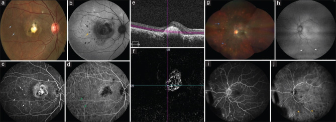
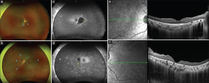
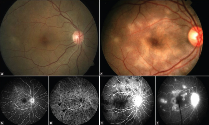
The white dot syndromes are a group of phenotypically similar disorders characterized by multiple lesions at the level of the outer retina, retinal pigment epithelium, and choroid. Common white dot syndromes whose imaging modalities have been described in this article are multiple evanescent white dot syndrome, acute posterior multifocal placoid pigment epitheliopathy, acute zonal occult outer retinopathy, multifocal choroiditis and panuveitis, punctate inner choroidopathy, serpiginous choroiditis, and birdshot chorioretinopathy. The various imaging modalities help us to better understand the pathophysiology of the various entities and help in diagnosing, monitoring, and prognosticating them. Optical coherence tomography angiography (OCTA) is a comparatively newer tool that helps us to visualize lesions in the choroid that correlate with indocyanine green angiography (ICGA) findings. Even though it is of limited value and cannot replace ICGA, it had gained considerable interest among ophthalmologists. Similarly, the noninvasive nature of modalities such as fundus autofluorescence and OCT makes them appealing and preferable over invasive techniques such as fundus fluorescein angiography and ICGA.
期刊介绍:
To provide a platform for scientific expression of the Oman Ophthalmic Society and the international Ophthalmic community and to provide opportunities for free exchange of ideas and information. To serve as a valuable resource for ophthalmologists, eye-care providers including optometrists, orthoptists, other health care professionals and research workers in all aspects of the field of visual science.


 分享
分享
 求助内容:
求助内容: 应助结果提醒方式:
 扫码关注我们
