{"title":"对侧眶外伤所致颈海绵状瘘。","authors":"V G Namitha, R Neena, E R Jayadevan","doi":"10.4103/ojo.ojo_258_23","DOIUrl":null,"url":null,"abstract":"<p><p>Direct carotid cavernous fistula is an abnormal arterio-venous connection from the carotid artery to the cavernous sinus (CS), resulting in high-pressure arterial blood entering the low-pressure venous CS. Most often, it occurs posttrauma and presents with ipsilateral orbital signs. In this report, we describe the case of a 54-year-old man, who presented with a late-onset right-sided red eye and diplopia following contralateral (left sided) orbital trauma (road traffic accident 7 months ago). Ocular examination revealed signs of Lateral rectus palsy, axial proptosis, and elevated intraocular pressure with dilated episcleral vessels in the right eye. Clinical findings were consistent with the diagnosis of right-sided direct CCF. To our surprise, digital subtraction angiogram revealed a left CCF with prominence of signs on the contralateral side. He underwent near-total endovascular coiling of the fistula, with initial aggravation of symptoms followed by near-total resolution.</p>","PeriodicalId":19461,"journal":{"name":"Oman Journal of Ophthalmology","volume":"17 3","pages":"384-387"},"PeriodicalIF":0.0000,"publicationDate":"2024-10-24","publicationTypes":"Journal Article","fieldsOfStudy":null,"isOpenAccess":false,"openAccessPdf":"https://www.ncbi.nlm.nih.gov/pmc/articles/PMC11620318/pdf/","citationCount":"0","resultStr":"{\"title\":\"Carotid-cavernous fistula due to contralateral orbital trauma.\",\"authors\":\"V G Namitha, R Neena, E R Jayadevan\",\"doi\":\"10.4103/ojo.ojo_258_23\",\"DOIUrl\":null,\"url\":null,\"abstract\":\"<p><p>Direct carotid cavernous fistula is an abnormal arterio-venous connection from the carotid artery to the cavernous sinus (CS), resulting in high-pressure arterial blood entering the low-pressure venous CS. Most often, it occurs posttrauma and presents with ipsilateral orbital signs. In this report, we describe the case of a 54-year-old man, who presented with a late-onset right-sided red eye and diplopia following contralateral (left sided) orbital trauma (road traffic accident 7 months ago). Ocular examination revealed signs of Lateral rectus palsy, axial proptosis, and elevated intraocular pressure with dilated episcleral vessels in the right eye. Clinical findings were consistent with the diagnosis of right-sided direct CCF. To our surprise, digital subtraction angiogram revealed a left CCF with prominence of signs on the contralateral side. He underwent near-total endovascular coiling of the fistula, with initial aggravation of symptoms followed by near-total resolution.</p>\",\"PeriodicalId\":19461,\"journal\":{\"name\":\"Oman Journal of Ophthalmology\",\"volume\":\"17 3\",\"pages\":\"384-387\"},\"PeriodicalIF\":0.0000,\"publicationDate\":\"2024-10-24\",\"publicationTypes\":\"Journal Article\",\"fieldsOfStudy\":null,\"isOpenAccess\":false,\"openAccessPdf\":\"https://www.ncbi.nlm.nih.gov/pmc/articles/PMC11620318/pdf/\",\"citationCount\":\"0\",\"resultStr\":null,\"platform\":\"Semanticscholar\",\"paperid\":null,\"PeriodicalName\":\"Oman Journal of Ophthalmology\",\"FirstCategoryId\":\"1085\",\"ListUrlMain\":\"https://doi.org/10.4103/ojo.ojo_258_23\",\"RegionNum\":0,\"RegionCategory\":null,\"ArticlePicture\":[],\"TitleCN\":null,\"AbstractTextCN\":null,\"PMCID\":null,\"EPubDate\":\"2024/9/1 0:00:00\",\"PubModel\":\"eCollection\",\"JCR\":\"Q3\",\"JCRName\":\"Medicine\",\"Score\":null,\"Total\":0}","platform":"Semanticscholar","paperid":null,"PeriodicalName":"Oman Journal of Ophthalmology","FirstCategoryId":"1085","ListUrlMain":"https://doi.org/10.4103/ojo.ojo_258_23","RegionNum":0,"RegionCategory":null,"ArticlePicture":[],"TitleCN":null,"AbstractTextCN":null,"PMCID":null,"EPubDate":"2024/9/1 0:00:00","PubModel":"eCollection","JCR":"Q3","JCRName":"Medicine","Score":null,"Total":0}
Carotid-cavernous fistula due to contralateral orbital trauma.
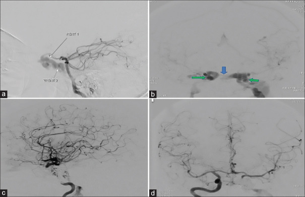
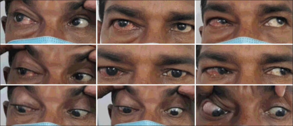
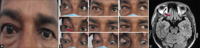
Direct carotid cavernous fistula is an abnormal arterio-venous connection from the carotid artery to the cavernous sinus (CS), resulting in high-pressure arterial blood entering the low-pressure venous CS. Most often, it occurs posttrauma and presents with ipsilateral orbital signs. In this report, we describe the case of a 54-year-old man, who presented with a late-onset right-sided red eye and diplopia following contralateral (left sided) orbital trauma (road traffic accident 7 months ago). Ocular examination revealed signs of Lateral rectus palsy, axial proptosis, and elevated intraocular pressure with dilated episcleral vessels in the right eye. Clinical findings were consistent with the diagnosis of right-sided direct CCF. To our surprise, digital subtraction angiogram revealed a left CCF with prominence of signs on the contralateral side. He underwent near-total endovascular coiling of the fistula, with initial aggravation of symptoms followed by near-total resolution.
期刊介绍:
To provide a platform for scientific expression of the Oman Ophthalmic Society and the international Ophthalmic community and to provide opportunities for free exchange of ideas and information. To serve as a valuable resource for ophthalmologists, eye-care providers including optometrists, orthoptists, other health care professionals and research workers in all aspects of the field of visual science.


 分享
分享
 求助内容:
求助内容: 应助结果提醒方式:
 扫码关注我们
