Maarten C Tol, Rick H W de Vries, Marten A Engelse, Françoise Carlotti, Aart A van Apeldoorn, Eelco J P de Koning, Volkert A L Huurman
{"title":"猪开放微孔胰岛输送装置的皮下植入。","authors":"Maarten C Tol, Rick H W de Vries, Marten A Engelse, Françoise Carlotti, Aart A van Apeldoorn, Eelco J P de Koning, Volkert A L Huurman","doi":"10.1177/15533506241306491","DOIUrl":null,"url":null,"abstract":"<p><p>BackgroundIntraportal pancreatic islet transplantation is a treatment option for patients with severe beta cell failure and unstable glycemic control. However, this procedure is associated with loss of beta cells after intrahepatic transplantation. Islet delivery devices (IDDs) implanted at extrahepatic sites may support engraftment and improve survival of pancreatic islets. We assessed the surgical feasibility, tolerability and safety of implantation of open microwell devices at subcutaneous sites with varying friction in pigs.MethodsOpen, non-immunoisolating microwell islet delivery devices were made from polyvinylidene fluoride (PVDF). Empty (n = 26) and islet-seeded devices (n = 8) were implanted subcutaneously in 6 immunocompetent pigs in low-friction sites (abdomen and lateral hip) and high-friction sites (anterior neck) for 3 months. Retrieved grafts were analyzed histologically with haematoxylin and eosin, and Masson's Trichrome staining.ResultsIslet-seeding and transportation of IDDs was free from complications with minimal islet spillage. IDDs were implanted subcutaneously using standard surgical equipment, without complications during the surgeries. IDDs implanted in the neck and IDDs co-transplanted with human islets were expelled and retrieved after 10 days. Empty IDDs were removed after 3 months. The abdominal site showed reduced signs of inflammation as compared to the neck region, while similar tissue ingrowth and vascularization of devices were found in the two locations.ConclusionsOpen microwell IDDs can safely be implanted with standard surgical equipment and successful islet-loading can be performed. Low-friction sites are preferable over high-friction sites for subcutaneous implantation in the porcine model since these lead to the least amount of foreign body reaction.</p>","PeriodicalId":22095,"journal":{"name":"Surgical Innovation","volume":" ","pages":"141-148"},"PeriodicalIF":2.2000,"publicationDate":"2025-04-01","publicationTypes":"Journal Article","fieldsOfStudy":null,"isOpenAccess":false,"openAccessPdf":"https://www.ncbi.nlm.nih.gov/pmc/articles/PMC11894865/pdf/","citationCount":"0","resultStr":"{\"title\":\"Subcutaneous Implantation of Open Microwell Islet Delivery Devices in Pigs.\",\"authors\":\"Maarten C Tol, Rick H W de Vries, Marten A Engelse, Françoise Carlotti, Aart A van Apeldoorn, Eelco J P de Koning, Volkert A L Huurman\",\"doi\":\"10.1177/15533506241306491\",\"DOIUrl\":null,\"url\":null,\"abstract\":\"<p><p>BackgroundIntraportal pancreatic islet transplantation is a treatment option for patients with severe beta cell failure and unstable glycemic control. However, this procedure is associated with loss of beta cells after intrahepatic transplantation. Islet delivery devices (IDDs) implanted at extrahepatic sites may support engraftment and improve survival of pancreatic islets. We assessed the surgical feasibility, tolerability and safety of implantation of open microwell devices at subcutaneous sites with varying friction in pigs.MethodsOpen, non-immunoisolating microwell islet delivery devices were made from polyvinylidene fluoride (PVDF). Empty (n = 26) and islet-seeded devices (n = 8) were implanted subcutaneously in 6 immunocompetent pigs in low-friction sites (abdomen and lateral hip) and high-friction sites (anterior neck) for 3 months. Retrieved grafts were analyzed histologically with haematoxylin and eosin, and Masson's Trichrome staining.ResultsIslet-seeding and transportation of IDDs was free from complications with minimal islet spillage. IDDs were implanted subcutaneously using standard surgical equipment, without complications during the surgeries. IDDs implanted in the neck and IDDs co-transplanted with human islets were expelled and retrieved after 10 days. Empty IDDs were removed after 3 months. The abdominal site showed reduced signs of inflammation as compared to the neck region, while similar tissue ingrowth and vascularization of devices were found in the two locations.ConclusionsOpen microwell IDDs can safely be implanted with standard surgical equipment and successful islet-loading can be performed. Low-friction sites are preferable over high-friction sites for subcutaneous implantation in the porcine model since these lead to the least amount of foreign body reaction.</p>\",\"PeriodicalId\":22095,\"journal\":{\"name\":\"Surgical Innovation\",\"volume\":\" \",\"pages\":\"141-148\"},\"PeriodicalIF\":2.2000,\"publicationDate\":\"2025-04-01\",\"publicationTypes\":\"Journal Article\",\"fieldsOfStudy\":null,\"isOpenAccess\":false,\"openAccessPdf\":\"https://www.ncbi.nlm.nih.gov/pmc/articles/PMC11894865/pdf/\",\"citationCount\":\"0\",\"resultStr\":null,\"platform\":\"Semanticscholar\",\"paperid\":null,\"PeriodicalName\":\"Surgical Innovation\",\"FirstCategoryId\":\"3\",\"ListUrlMain\":\"https://doi.org/10.1177/15533506241306491\",\"RegionNum\":4,\"RegionCategory\":\"医学\",\"ArticlePicture\":[],\"TitleCN\":null,\"AbstractTextCN\":null,\"PMCID\":null,\"EPubDate\":\"2024/12/13 0:00:00\",\"PubModel\":\"Epub\",\"JCR\":\"Q3\",\"JCRName\":\"SURGERY\",\"Score\":null,\"Total\":0}","platform":"Semanticscholar","paperid":null,"PeriodicalName":"Surgical Innovation","FirstCategoryId":"3","ListUrlMain":"https://doi.org/10.1177/15533506241306491","RegionNum":4,"RegionCategory":"医学","ArticlePicture":[],"TitleCN":null,"AbstractTextCN":null,"PMCID":null,"EPubDate":"2024/12/13 0:00:00","PubModel":"Epub","JCR":"Q3","JCRName":"SURGERY","Score":null,"Total":0}
Subcutaneous Implantation of Open Microwell Islet Delivery Devices in Pigs.
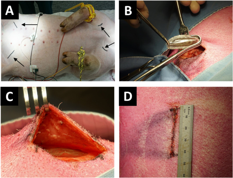
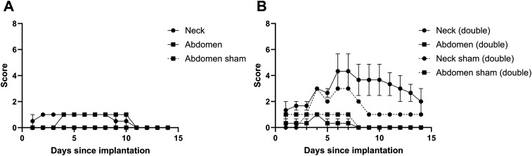
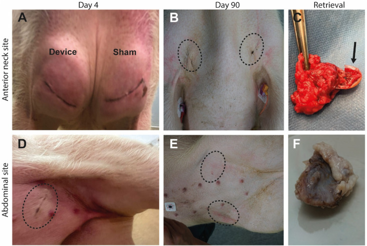
BackgroundIntraportal pancreatic islet transplantation is a treatment option for patients with severe beta cell failure and unstable glycemic control. However, this procedure is associated with loss of beta cells after intrahepatic transplantation. Islet delivery devices (IDDs) implanted at extrahepatic sites may support engraftment and improve survival of pancreatic islets. We assessed the surgical feasibility, tolerability and safety of implantation of open microwell devices at subcutaneous sites with varying friction in pigs.MethodsOpen, non-immunoisolating microwell islet delivery devices were made from polyvinylidene fluoride (PVDF). Empty (n = 26) and islet-seeded devices (n = 8) were implanted subcutaneously in 6 immunocompetent pigs in low-friction sites (abdomen and lateral hip) and high-friction sites (anterior neck) for 3 months. Retrieved grafts were analyzed histologically with haematoxylin and eosin, and Masson's Trichrome staining.ResultsIslet-seeding and transportation of IDDs was free from complications with minimal islet spillage. IDDs were implanted subcutaneously using standard surgical equipment, without complications during the surgeries. IDDs implanted in the neck and IDDs co-transplanted with human islets were expelled and retrieved after 10 days. Empty IDDs were removed after 3 months. The abdominal site showed reduced signs of inflammation as compared to the neck region, while similar tissue ingrowth and vascularization of devices were found in the two locations.ConclusionsOpen microwell IDDs can safely be implanted with standard surgical equipment and successful islet-loading can be performed. Low-friction sites are preferable over high-friction sites for subcutaneous implantation in the porcine model since these lead to the least amount of foreign body reaction.
期刊介绍:
Surgical Innovation (SRI) is a peer-reviewed bi-monthly journal focusing on minimally invasive surgical techniques, new instruments such as laparoscopes and endoscopes, and new technologies. SRI prepares surgeons to think and work in "the operating room of the future" through learning new techniques, understanding and adapting to new technologies, maintaining surgical competencies, and applying surgical outcomes data to their practices. This journal is a member of the Committee on Publication Ethics (COPE).


 分享
分享
 求助内容:
求助内容: 应助结果提醒方式:
 扫码关注我们
