{"title":"猫脑内良性脑膜瘤的侵袭。","authors":"Kiyotaka Arai, Shinichi Nakamura, Kanami Matsubara, Yuma Ozaki, Sho Kadekaru, Akihiko Sugiyama, Ikki Mitsui, Natsuki Akashi, Ryohei Yoshitake, Kenji Kutara","doi":"10.1177/20551169241291842","DOIUrl":null,"url":null,"abstract":"<p><strong>Case summary: </strong>A 13-year-old castrated male American Shorthair cat was referred for evaluation following a 3-week history of poor balance and decreased activity. The MRI findings revealed a well-defined left caudal cerebellar mass with a diameter of 1.2 cm, consistent with a meningioma. CT and MRI scans did not reveal metastasis. After a suboccipital craniotomy, the mass was resected subtotally as a result of parenchymal invasion. A histopathological examination revealed a benign fibrous meningioma with invasion into the cerebellum at the tumour margins. Postoperatively, the cat remained asymptomatic for 25 months, but ataxia recurred, and tumour recurrence was confirmed using MRI and post-mortem examination.</p><p><strong>Relevance and novel information: </strong>This case demonstrates that even feline meningiomas with benign features can exhibit brain invasion. Since brain invasion is an indicator of malignancy in meningioma, a detailed histopathological evaluation of the tumour margins is essential for accurate grading and prognosis.</p>","PeriodicalId":36588,"journal":{"name":"Journal of Feline Medicine and Surgery Open Reports","volume":"10 2","pages":"20551169241291842"},"PeriodicalIF":0.7000,"publicationDate":"2024-12-16","publicationTypes":"Journal Article","fieldsOfStudy":null,"isOpenAccess":false,"openAccessPdf":"https://www.ncbi.nlm.nih.gov/pmc/articles/PMC11650476/pdf/","citationCount":"0","resultStr":"{\"title\":\"Brain invasion by an otherwise benign meningioma in a cat.\",\"authors\":\"Kiyotaka Arai, Shinichi Nakamura, Kanami Matsubara, Yuma Ozaki, Sho Kadekaru, Akihiko Sugiyama, Ikki Mitsui, Natsuki Akashi, Ryohei Yoshitake, Kenji Kutara\",\"doi\":\"10.1177/20551169241291842\",\"DOIUrl\":null,\"url\":null,\"abstract\":\"<p><strong>Case summary: </strong>A 13-year-old castrated male American Shorthair cat was referred for evaluation following a 3-week history of poor balance and decreased activity. The MRI findings revealed a well-defined left caudal cerebellar mass with a diameter of 1.2 cm, consistent with a meningioma. CT and MRI scans did not reveal metastasis. After a suboccipital craniotomy, the mass was resected subtotally as a result of parenchymal invasion. A histopathological examination revealed a benign fibrous meningioma with invasion into the cerebellum at the tumour margins. Postoperatively, the cat remained asymptomatic for 25 months, but ataxia recurred, and tumour recurrence was confirmed using MRI and post-mortem examination.</p><p><strong>Relevance and novel information: </strong>This case demonstrates that even feline meningiomas with benign features can exhibit brain invasion. Since brain invasion is an indicator of malignancy in meningioma, a detailed histopathological evaluation of the tumour margins is essential for accurate grading and prognosis.</p>\",\"PeriodicalId\":36588,\"journal\":{\"name\":\"Journal of Feline Medicine and Surgery Open Reports\",\"volume\":\"10 2\",\"pages\":\"20551169241291842\"},\"PeriodicalIF\":0.7000,\"publicationDate\":\"2024-12-16\",\"publicationTypes\":\"Journal Article\",\"fieldsOfStudy\":null,\"isOpenAccess\":false,\"openAccessPdf\":\"https://www.ncbi.nlm.nih.gov/pmc/articles/PMC11650476/pdf/\",\"citationCount\":\"0\",\"resultStr\":null,\"platform\":\"Semanticscholar\",\"paperid\":null,\"PeriodicalName\":\"Journal of Feline Medicine and Surgery Open Reports\",\"FirstCategoryId\":\"1085\",\"ListUrlMain\":\"https://doi.org/10.1177/20551169241291842\",\"RegionNum\":0,\"RegionCategory\":null,\"ArticlePicture\":[],\"TitleCN\":null,\"AbstractTextCN\":null,\"PMCID\":null,\"EPubDate\":\"2024/7/1 0:00:00\",\"PubModel\":\"eCollection\",\"JCR\":\"Q3\",\"JCRName\":\"VETERINARY SCIENCES\",\"Score\":null,\"Total\":0}","platform":"Semanticscholar","paperid":null,"PeriodicalName":"Journal of Feline Medicine and Surgery Open Reports","FirstCategoryId":"1085","ListUrlMain":"https://doi.org/10.1177/20551169241291842","RegionNum":0,"RegionCategory":null,"ArticlePicture":[],"TitleCN":null,"AbstractTextCN":null,"PMCID":null,"EPubDate":"2024/7/1 0:00:00","PubModel":"eCollection","JCR":"Q3","JCRName":"VETERINARY SCIENCES","Score":null,"Total":0}
Brain invasion by an otherwise benign meningioma in a cat.
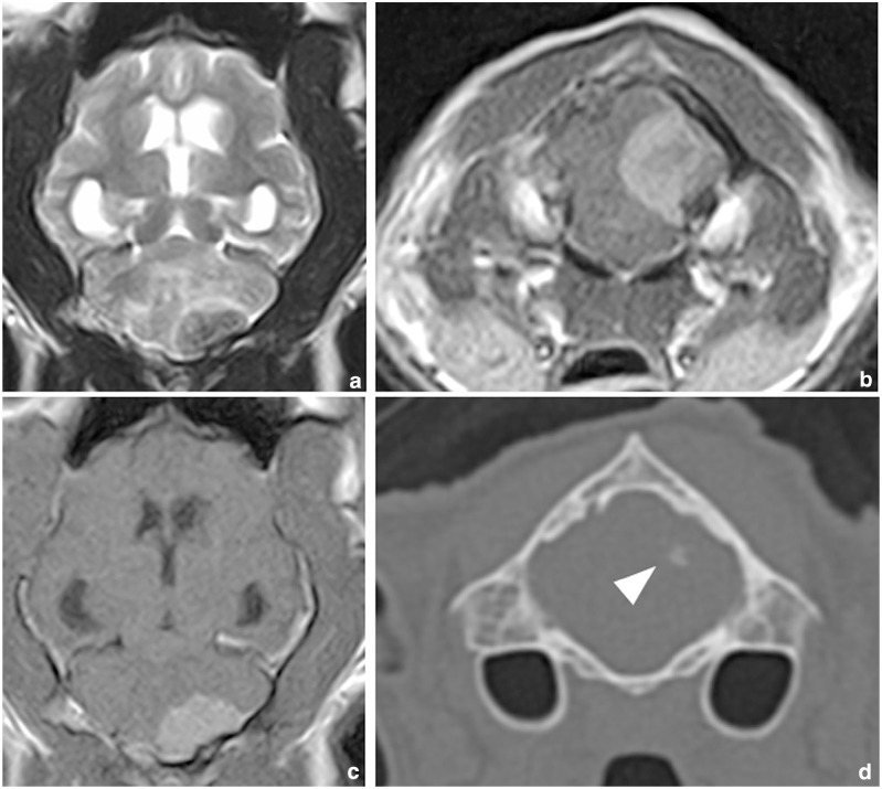
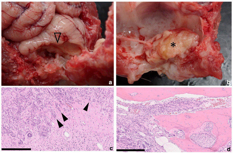
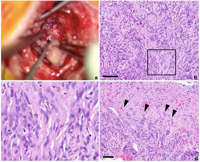
Case summary: A 13-year-old castrated male American Shorthair cat was referred for evaluation following a 3-week history of poor balance and decreased activity. The MRI findings revealed a well-defined left caudal cerebellar mass with a diameter of 1.2 cm, consistent with a meningioma. CT and MRI scans did not reveal metastasis. After a suboccipital craniotomy, the mass was resected subtotally as a result of parenchymal invasion. A histopathological examination revealed a benign fibrous meningioma with invasion into the cerebellum at the tumour margins. Postoperatively, the cat remained asymptomatic for 25 months, but ataxia recurred, and tumour recurrence was confirmed using MRI and post-mortem examination.
Relevance and novel information: This case demonstrates that even feline meningiomas with benign features can exhibit brain invasion. Since brain invasion is an indicator of malignancy in meningioma, a detailed histopathological evaluation of the tumour margins is essential for accurate grading and prognosis.


 分享
分享
 求助内容:
求助内容: 应助结果提醒方式:
 扫码关注我们
